


INTRODUCTION
Hepatitis C virus (HCV) is a major human hepatotropic pathogen that accounts for a significant global disease burden. Approximately 50 million people worldwide are infected with chronic Hepatitis C, with an annual rate of over one million new infections. The alarming number was first red-flagged by the WHO, which cited 242,000 deaths in 2022 due to HCV-related hepatopathies. The global death rates due to HCV infection ranked from 10th to 7th place from 1990 to 2013 (1).
Early HCV infection is usually asymptomatic and, if untreated, gradually progresses to chronic illness primarily involving hepatotropism and lymphotropism. Mortality due to extrahepatic pathologies is double in HCV patients compared to non-infected individuals. Likewise, the death rates due to hepatic and extrahepatic pathologies are 10-fold higher in virus-infected patients than in non-infected, suggesting the systemic nature of viral infection (2).
Strategic remedies include effective anti-HCV treatment with DAAs that inhibit the non-structural viral replication proteins, namely NS3/4A serine protease, NS5A replication complex, and NS5B RNA-dependent RNA polymerase. Despite significantly higher virological cure rates (>95%), the asymptomatic nature of the disease, high treatment costs, poor diagnostic strategies, RASs, low cirrhotic efficacy, drug-drug interactions, and pharmacokinetic variability are major challenges for current DAA therapy. However, a combinatorial therapeutic regimen has improved overall efficacy with subsided RAS effects (3). The development of pan-genotypic regimens with maximum effectiveness, shorter course duration, and minimal cross-drug interactions could mitigate downstream effects.
The virus acquired resistance against DAAs, transmission of resistant HCV variants and incidences of reinfection are paramount challenges for the effective eradication of disease. Moreover, there is evidence of post-infection long-term health complications, such as the progression of liver cirrhosis into cancer. Thus, the development of preventive vaccines will be a crucial approach for global control of HCV-induced chronic hepatopathies. However, the extraordinary genetic heterogeneity of HCV genotypes and subtypes, as well as their ability to evade host immunity, have hindered the development of a vaccine. At this time, rich literature is available that flags the mosaic information and inconclusiveness of the study.
In this study, we analyzed the molecular, phylogenetic, and immunological aspects of the HCV genome, highlighting the associated pathologies and global epidemiology, as well as the progress and challenges in HCV vaccine development.
MOLECULAR TAPESTRY OF HCV
HCV is a noncytopathic, blood-borne, enveloped virus. Its genome is a 9,600-nucleotide-long positive-sense single- stranded RNA, delimited by conserved untranslated regions (UTRs). The in-frame single ORF flanked by the 5'- and 3′-UTR encodes a polyprotein composed of structural and non-structural proteins (Fig. 1). While the former makes up the viral envelope and nucleocapsid, the latter are largely viral replication proteins. The 5'-UTR exhibits translational control, and the 3'-UTR regulates viral replication (4, 5).
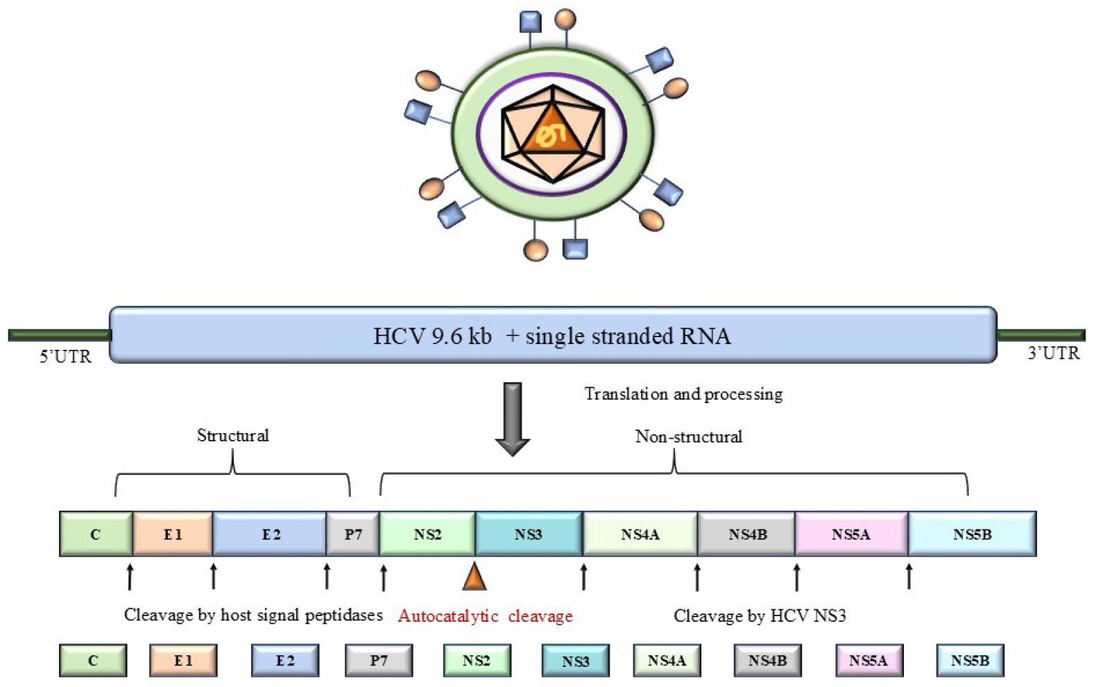
The 5′-UTR is the most conserved region upstream of the core protein. It folds into a modular structure made of four distinct secondary structural elements designated as domains I-IV. Domains II, III, and IV constitute the Internal Ribosome Entry Site (IRES) essential for atypical cap-independent translation initiation of the viral RNA. The complex three- dimensional folds in domains II and III are critical for interactions with the 40S ribosomal subunit. Domain IV comprises a start codon that melts into a linear single-stranded mRNA, placing the 40S ribosome subunit with initiator tRNA directly over the start codon, thereby skipping the 5'-cap scanning process. Domain I is not part of IRES, although it is docked by host liver-specific miRNA-122, a proviral factor with protective and regulatory roles during viral replication and translation.
The 3′-UTR has three distinct regions: a variable region of 30-40 nucleotides, a 90-nucleotide-long poly (U/UC) spacer, and an invariable 3′-X tail of 98 nucleotides, which attains a three- or two-stem-loop conformation. The formation of the loop exposes a dimer linkage sequence, which contributes to the dimerization of viral RNA in vitro. Since dimerization can affect HCV replication, this might represent an intermediate conformation while switching between viral replication and encapsidation. This region probably engages with the 5'-UTR to stabilize RNA for enhanced translational efficiency. The 3′-UTR makes distant regulatory interactions with several RNA elements present within coding regions and serves as an intrinsic cis-regulatory element (CRE) during viral replication and translation. One such CRE is present in the NS5B coding region. The 3′-X tail serves as a promoter where NS5B binds to synthesize complementary (-)ssRNA. The poly U/UC stretch is crucial for replication complex assembly, providing conformational flexibility to the downstream stem-loops to facilitate distant interactions with CRE (6).
The core comprises RNA and lipid-binding nucleocapsid-forming structural proteins produced as a two-step cleavage product. The initial cleavage by host signal peptidase yields a 191 residues long precursor, followed by a second signal peptide peptidase-mediated cleavage that removes the C-terminal E1 signal sequence to produce a mature 177 aa-long core protein. The mature core protein has an N-terminal D1 domain and a C-terminal D2 (Fig. 2A). D1 is a flexible structure comprising three basic subdomains: BD1, BD2, and BD3, which bind to viral RNA and use it as a scaffold for oligomerization during nucleocapsid packaging. The adjoining D2 domain has two helices (H1 and H2) separated by a hydrophobic loop tethering the core protein to the ER membrane and lipid droplets. The extreme C-terminal stretch of 20 residues constitutes the D3 domain, which is involved in the modulation of multiple host proteins, cell cycle and signaling pathways, lipid metabolism, and apoptosis. Interestingly, the core protein displays pro-apoptotic and/or anti-apoptotic properties in various cellular contexts. It promotes apoptosis of infected liver cells, which leads to hepatic injury and inflammation, while suppressing apoptosis for self-sustenance and immune evasion, thus leading to hepatocarcinoma (7, 8).
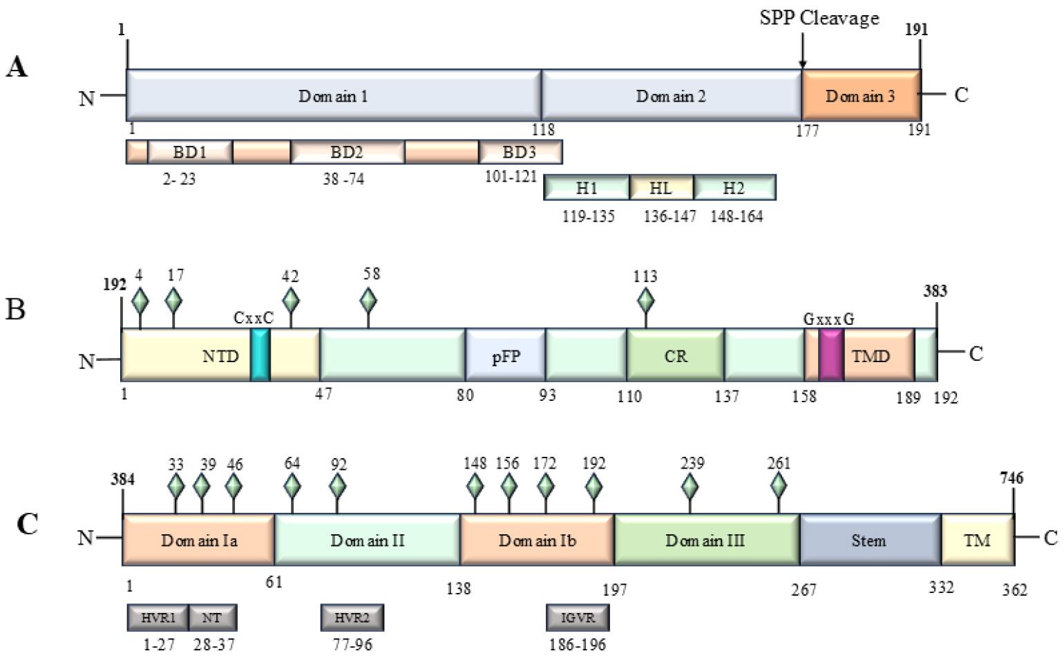
Fig. 2
Domain architecture of structural proteins. A. core protein; B. E1 glycoprotein; C. E2 glycoprotein.
E1 and E2 are structural type 1 transmembrane glycoproteins of the HCV envelope that exist as obligate non-covalent heterodimers. The ectodomain of E1 possesses an N-terminal domain (NTD), putative fusion peptide (pFP), and conserved region (CR). The E2 ectodomain has two hypervariable regions (HVR1 and HVR2), followed by an intergenic variable region (IGVR) and a stem region. E1/E2 heterodimer is critical for viral entry and fusion with the host cell membrane (9). The membrane-spanning regions of E1 and E2 contain two α-helices separated by a short stretch of basic residues (GPRL motif in E1 and R/KGY motif in E2) are conserved across all genotypes. It facilitates E1/E2 heterodimerization, ER localization, membrane anchoring, and ER retention (10, 11, 12). E1 and E2 proteins are heavily glycosylated with up to five and eleven N-glycosylation sites, respectively (Fig. 2B and Fig. 2C). The four N-linked glycosylation sites (Asn-196, 209, 234, and 305), which are key to functional E1 folding and heterodimerization, are conserved across all genotypes, whereas the fifth site (Asn-250) is restricted to GT1b and GT6 (13). The glycans of E2 are generally conserved. However, exceptions like the absence of N476 in GT1 and GT2, have been reported. The glycans of E1 get masked upon heterodimerization, whereas the glycans of E2 possess immunodominant neutralizing epitopes acting as immunological decoys for more conserved neutralizing epitopes. It was probably evolved as a robust way to evade host immunity. HVRs display extreme sequence variability (~80%) and regulate their overall binding affinity with CD81 receptors, thereby dictating the strength of the E2-receptor complex for different genotypes (14). The inevitable similarity recorded in the 3D conformations and physicochemical properties of E2 glycoproteins conserves the function of initial host cell recognition and attachment during viral entry (15). The E1/E2 heterodimer complements each other in host receptor attachment, endocytosis, and endosomal membrane fusion during viral entry.
The p7 is a small (7 kDa) hydrophobic protein that contributes to the packaging, budding, and release of viral particles (Fig. 3A). It has two α-helical transmembrane domains separated by a cytoplasmic loop with both termini facing the ER lumen of the cell. The p7 protein is a viroporin that oligomerizes as hepta- or hexamers to form cation channels, altering the permeability of the host membrane and modulating the pH of secretory compartments to prevent the inactivation of viral glycoproteins (16, 17). The cytoplasmic loop is important for viral pathogenesis. The intraluminal regions of p7 possess genotype-based functionality, as shown by the loss-of-function in GT1a chimeras, wherein the p7 region was swapped with the GT2a sequence (18).
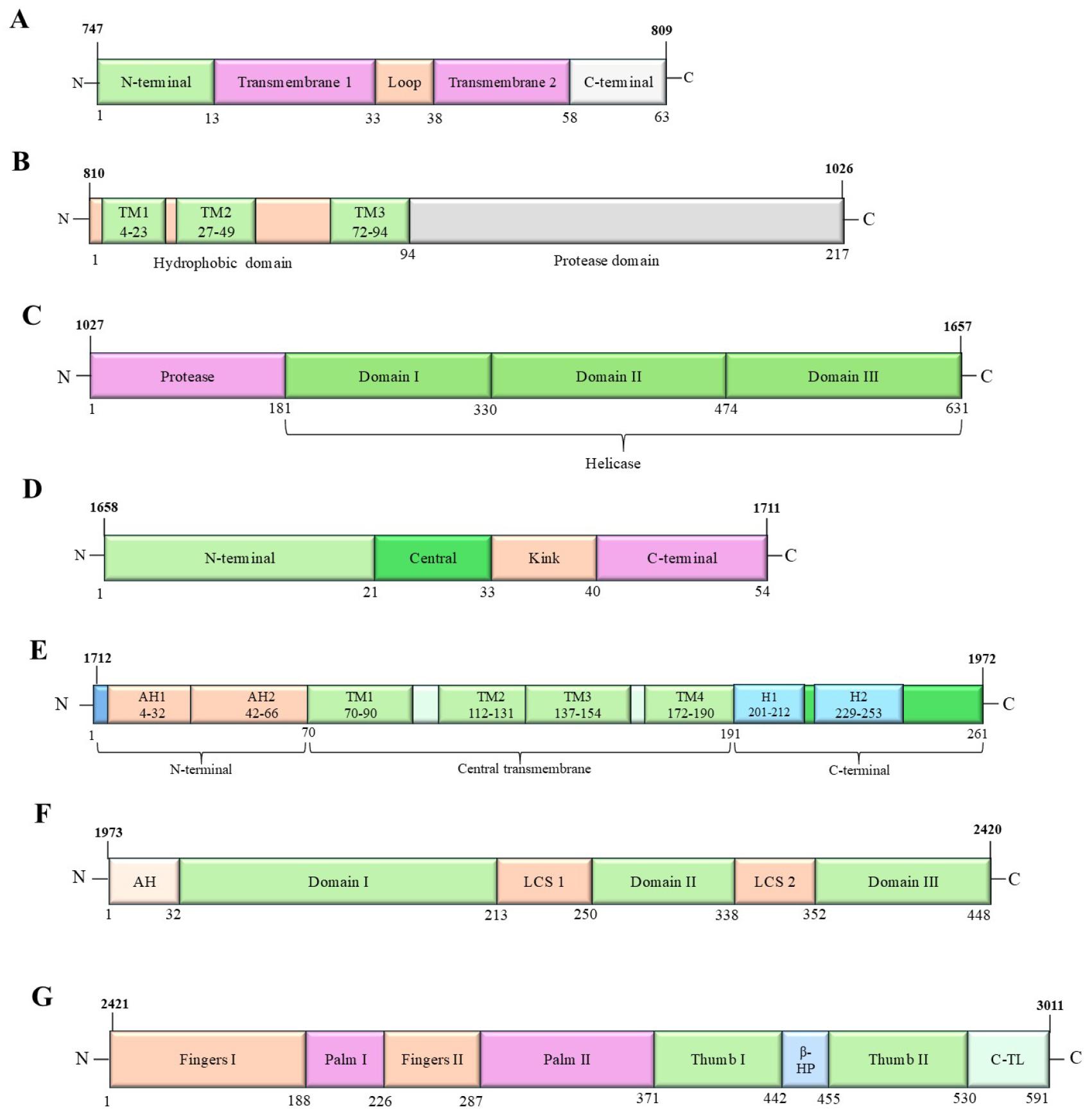
Fig. 3
Domain architecture of non-structural proteins. A. p7 protein; B. NS2 protein; C. NS3 protein; D. NS4A protein; E. NS4B protein; F. NS5A protein; G. NS5B protein (RdRP).
NS2 is a non-structural dual-function integral membrane protein that functions as cysteine protease and a scaffolding factor during viral assembly (Fig. 3B). It has internal signal sequences (839-883 and 928-960 residues) that facilitate co-translational translocation of NS2 in the ER membrane (19). The N-terminus has three TM domains for ER-membrane integration, while the cytosolic C-terminal is a globular protease, which, along with the N- terminus of NS3, functions as a Zn-dependent metalloprotease to cleave NS2-NS3 junction (20). This proteolysis also requires an ER-resident signal recognition particle receptor protein (21). In addition to the auto-proteolytic activity, NS2 is vital for the recruitment of E2, p7, NS3, and NS5A in the vicinity of LDs for their final assembly into virions (22).
NS3 is a 70-kDa dual-function enzyme with an N-terminal (180 aa) serine protease, and a C-terminal (450 aa) NTPase/ Helicase (Fig. 3C). The protease active site is a conserved triad of His57, Asp81 and Ser139. The NS3-NS4A enzyme complex can be a therapeutic target as it cleaves all the downstream non-structural viral proteins (NS3/NS4A, NS4A/NS4B, NS4B/NS5A, and NS5A/NS5B). Further, it proteolytically inactivates several host proteins that may interfere with viral replication (23). NS3 is a helicase that catalyzes the NTP-driven unwinding of RNA secondary structures and dsRNA intermediates during viral replication (24). Moreover, NS3 is important for NS5B-mediated template recognition in the replication complex.
NS4A is a cofactor that facilitates genome replication and viral assembly through NS4B and NS3, respectively. Its membrane-spanning N-terminal helix integrates the NS3-NS4A complex into the membrane, whereas the central region contributes to NS3 folding. The C-terminus has a highly acidic stretch that regulates NS5A hyperphosphorylation (Fig. 3D).
NS4B is an integral transmembrane protein with a complex topology localized in the ER. Its amphipathic N-terminal helix faces the cytoplasm, which, upon post-translational cleavage, flips into the ER lumen. This dual topology facilitates the modulation of the ER membrane to form a membranous web that supports replication complex assembly and shields the genome from immune detection. The middle part is composed of four transmembrane helices, followed by a helical C-terminus in the cytosol (25, 26) (Fig. 3E). NS4A and NS4B are the only translational inhibitors of all the non-structural viral proteins, which not only close down the cellular protein translation but also retard the IRES-mediated viral translation. This ensures the long-term persistence of the virus in the host (27, 28).
NS5A, a Zn-dependent metallophosphoprotein, is essential for the formation of ER-derived double-membrane vesicles for the organization of the virus replication machinery. The initial 30 N-terminus residues form an amphipathic α-helix that anchors the protein to the cytoplasmic leaflet of the ER membrane. The following D1 domain is a conserved, dimeric RNA-binding structure with Zn-binding motifs and attains a basal-phosphorylation state that is essential for replication. The D2 and D3 domains are intrinsically disordered. These three domains are separated by two low-complexity sequences (LCS1 and LCS2), which, when phosphorylated, switch the protein function involving domain D3. Initially, NS5A localizes to the membranous web and contributes to RC establishment and replication efficiency. During the late stages of the viral life cycle and in the hyperphosphorylated state, it co-localizes with core and NS2 proteins to LDs and creates a scaffold for virion assembly. Moreover, it antagonistically interacts with host cellular proteins, particularly IFN signaling components, to ensure the chronic persistence of the virus within hepatocytes. Intervention with components of growth-related pathways progressively leads to HCC (Fig. 3F).
NS5B is a viral RNA-dependent RNA polymerase (RdRp) with a typical right-hand topology (Fig. 3G). It has a C-terminal single-pass, helical transmembrane domain for membrane insertion. Post-translational targeting of NS5B to the cytosolic side of the ER membrane exposes its functional polymerase domain (29). It dwells between closed and open conformations during nucleotide incorporation and subsequent template elongation. An unusually extensive interaction between the thumb and extensions of the finger (fingertips) subdomain completely encircles the active site in the palm domain (30), supporting the unique de novo mechanism of RNA synthesis by viral RdRp. The enzyme also undergoes an unusual conformational change when switching from the initiation to the elongation phase. X-ray crystallography structures elucidating the active and allosteric sites of the enzyme provide critical insights for the development of structure-guided potential RdRp inhibitors (31, 32). The molecular characteristics and therapeutic potential (33, 34, 35, 36, 37, 38, 39, 40, 41, 42, 43, 44) of HCV proteins are summarized in Table 1.
Table 1.
Summary of molecular characteristic features and therapeutic potential of HCV proteins (ID AAA72945)
|
Viral proteins (aa range) |
Structural Aspect | Interaction Partners | PTM /processing | Role of viral protein | Therapeutic potential | |
|---|---|---|---|---|---|---|
| Viral proteins | Host factors | Modified residues | ||||
|
Core (p21: 2-177) (p23: 2-191) | Homo -oligomer | E1, NS5A | STAT1, STAT3, LTBR receptor, TNFRSF1A, HNRPK, YWHAE, UBE3A, DDX3X, APOA2, RXRA, SP110 isoform 3/Sp110b, CREB3, ACY3, C1QR1, RBM24, EIF2AK2/PKR |
N-acetylation (S2) Phosphorylation (S53) Phosphorylation (S99) Phosphorylation (S116) | Viral RNA packaging, Virion budding; RNA chaperon, Block IFN-α/β and IFN-γ signaling to inhibit antiviral state establishment in host-infected cells; Downregulate T-cell proliferation; Affects lipid metabolism; Increase triglyceride accumulation | HCVcAg can be used as a simple measure of active infection in DAA treatment monitoring (33) Possible target of plant-derived compounds like sylverin and naringenin (34, 35); Full-length Core protein recombinant vaccine targets cell-mediated immune response, in phase I clinical trial (36) |
|
E1 protein (192-383) | Heterodimer |
E2 Mature core protein |
N-glycosylation (N196) N-glycosylation (N209) N-glycosylation (N234) N-glycosylation (N305) |
Virus attachment, internalization, and fusion E1/E2 Form lipo-viro-particle (interaction with host apolipoproteins) |
Role in immune evasion; B-cell vaccine candidate; Mice immunized with the sE1 mRNA-LNP vaccine developed resistance against the surrogate vaccinia virus; Recombinant sE1/modified E2 mRNA LNP vaccine displayed improved immune response (37) | |
|
E2 protein (384-746) | Heterodimer |
E1 NS2 protease | CD81, SR-B1, EIF2AK2/PKR, CD209/DC-SIGN and CLEC4M/DC-SIGNR, SPCS1, SLC3A2/4F2hc, PLSCR1 |
O-glycosylation (T385) O-glycosylation (T396) O-glycosylation (S401) O-glycosylation (S404) N-glycosylation (N417) N-glycosylation (N423) Disulfide bond (C429-C552) N-glycosylation (N430) N-glycosylation (N448) Disulfide bond (C452-C459) O-glycosylation (T473) Disulfide bond (C486-C494) Disulfide bond (C503-508) O-glycosylation (T518) N-glycosylation (N532) N-glycosylation (N540) N-glycosylation (N556) Disulfide bond (C564-C569) N-glycosylation (N576) Disulfide bond (C581-C585) Disulfide bond (C597-C620) Disulfide bond (C607-C644) N-glycosylation (N623) N-glycosylation (N645) Disulfide bond (C652-C677) |
Virus attachment, internalization, and fusion E1/E2 form Lipo-viro-particle (interaction with host apolipoproteins) Inhibit the host's antiviral establishment |
Primary target of the B-cell vaccine; Role in immune evasion; Full-length recombinant E1/ E2 glycoproteins (HCV GT1a -clone H77) elicited improved antibody response and high antibody avidity (>75), in phase I human clinical trial (38); Modified E2 mRNA LNP demonstrated reduced CD81 binding, improved nAb response, and boosted total IgG level (37) |
|
P7 (747-809) |
Homo -hexamer Homo -heptamer | Protease NS2 |
Virion assembly, and release; An imbalanced ionic concentration in the host cytoplasm, an ion channel Mitochondrial depolarization Lipid raft | Potential role in immune evasion; Induce a polyfunctional cell-mediated immunity; target of inhibitors like amantadine, BIT225 and hexamethylene amiloride (39) | ||
|
NS2 Protease (810-1026) |
E/ E2, Viroporin p7 Serine protease/ Helicase NS3 | SPCS1 | S-palmitoylation (C922) | Viral particle assembly Autocleavage between NS2 and NS3 | No approved DAA | |
|
NS3 Serine protease /Helicase (1027-1657) | Protease NS2 NS4A | MAVS, TICAM1, TANK-binding kinase/TBK1, RBM24 |
Helicase, NTPase, and Serine protease activity Block the establishment of the antiviral state in infected hosT-cells. |
Quercetin, a NS3 protease inhibitor (40) CHRONAVACC (NS3/NS4 DNA vaccine induce T-cell response in phase I/IIa human clinical trial (41); Target for DAAs (3) | ||
|
NS4A (1658-1711) | NS3 Serine protease NS5A |
HCV propagation Cofactor for Serine protease NS3 |
Used in conjunction with NS3 as a DNA vaccine; Target of DAAs (3) | |||
|
NS4B (1712-1972) | Homo -multimer | NS5A | PLA2G4C, STING |
S-palmitoylation (C1968) S-palmitoylation (C1972) |
Inhibit IFN signaling Role in ER-derived membranous web formation | Inhibited by clemizole (42); PTC725 (43), 2-(4-sulfonamidophenyl)-indole 3-carboxamides (a promising pan-genotypic inhibitors (44); T-cell vaccine target |
|
NS5A (1973-2419) |
Monomer Homo-dimer |
NS4A NS4B RdRP | GRB2, BIN1, PIK3R1, SRCAP, FKBP8, VAPB, EIF2AK2/PKR, PACSIN2, SRC kinase, IFI27 and SKP2, GPS2, TNFRSF21, CIDEB, CHKA/choline kinase-α, SPB2, RAB18, PPIA/CYPA, TRIM 14 |
Phosphorylation in p56 (S2194) Phosphorylation in p58 (S2197) Phosphorylation in p58 (S2201) Phosphorylation in p58 (S2204) Phosphorylation in p58 (S2207) Phosphorylation in p58 (S2210) Cross-link (glycyl-lysine isopeptide, Gly-Lys2350) | Viral replication and assembly; Downregulation of IRES-dependent translation initiation; Prevent BIN1-induced apoptosis; Virus propagation; Modulation of lipid droplet formation | DAAs targeting NS5A include daclatasvir, ledipasvir, ombitasvir, elbasvir and velpatasvir (3) |
|
NS5B/ RdRP (2420-3010) |
Homo -oligomer | NS5A |
VAPB, PRK2/PKN2 HNRNPA1 and SEPT6 |
Phosphorylation (S2448)
Phosphor ylation (S2461) | Primer template recognition and RNA synthesis; Evasion of host antiviral machinery (5′ FAD capping) |
Luteolin a possible inhibitor (45); Sofosbuvir (NS5B nucleotide inhibitor) and dasabuvir (non-nucleoside polymerase inhibitor) (3); T-cell vaccine target |
LIFE CYCLE OF HCV
HCV particles are usually associated with lipoproteins and circulate in the bloodstream. Asymptomatic acute infection and ineffective clearance result in the progression of infection to hepatic cells. Liver sinusoidal endothelial cells (LSEC) and neighboring Kupffer cells expressing L-SIGN and DC-SIGN receptors, respectively, are the first to capture HCV virions circulating in the blood. Thereafter, viral particles bind to low-affinity attachment factors, such as heparin sulphate glycosaminoglycans and very low-density lipoprotein receptors found on the basolateral hepatocellular membranes. This concentrates the virions around hepatocytes, wherein high-affinity interactions with specific cell surface receptors (CD81 and SR-B1) propel HCV-receptor complexes to proximal tight junctions. The tight junction proteins, Claudin-1 and Occludin, together with CD81, initiate clathrin-mediated endocytic internalization of the virus-receptor complex which is a critical step in HCV genotype/subtype recognition. Post-internalization acidification of endosomes carrying viral entities causes fusion of viral and host membranes, thereby releasing the core viral particles into the cytoplasm (46).
Subsequent proteolysis of nucleocapsid proteins releases (+)ssRNA into the cytoplasm, which serves as an immediate template for translation. The intricate tertiary folds formed by the stem-loop secondary structure of the 5'-UTR-localized IRES serve as a direct docking site for the 40S ribosome. The resulting binary complex localizes to the rough ER, where viral mRNA translation and replication proceed through host machinery. Further, the modification of viral proteins, packaging and maturation of progeny virions, and their loading onto the host secretory pathway complete the life cycle of HCV.
Viral translation follows a cap-independent mechanism of translational initiation. HCV can exhibit simpler modes of translation initiation under stress and high cation levels (47). The translation product is a 3010 aa-long precursor polyprotein, which is cleaved by various host and viral proteases in a protein-specific manner to generate three structural (core, E1, and E2 glycoproteins) and seven non-structural proteins (P7, NS2, NS3, NS4A, NS4B, NS5A, and NS5B).
In contrast, viral replication is a complex process that is assisted by several host factors. An ER-derived aggregate of double- membrane vesicles, the membranous web, is the primary site for the association of viral replicative machinery. Liver-specific miRNA-122 binds the 5'UTR upstream of IRES as an Ago2:miRNA122 complex at two tandem sites, the S1 site near the stem-loop 1 with higher affinity and the S2 site at the base of stem loop 2, forming a 3′- overhang on the 5'- end of viral RNA. Upstream-bound miRNA-122 enables IRES to gain a translation-competent conformation, stabilizing genomic RNA from the 5'-exoribonuclease activity of Xrn1. The association of miRNA-122 at the S2 is competed by a host poly(C) binding protein 2 (PCBP2), serving as a translation-to-replication switch. While PCBP2-mediated circularization of genomic RNA enhances translational efficiency, miRNA-122 binding promotes viral replication (48). Although the impetus of miRNA-122 binding favors viral replication, it triggers immediate viral translation post-infection. In silico studies have predicted additional miRNA-122 binding sites in the viral genome: four in the NS5B coding region and three in the 3′-UTR, with limited information about their functional significance (49).
The virus uses fatty acid metabolism and the synthesis of very low-density lipoproteins for the assembly and release of new viral particles. The NS2 protein forms a complex with E1/E2, p7, and NS3 and NS5A to bridge the replicase machinery at the site of assembly of the viral progenies. Initially, viral RNA is transported through a network of NS5A proteins to cytoplasmic LDs coated with core proteins. The encapsidation of the viral genome occurs near ER-derived membranes, where E1/E2 glycoproteins accumulate with NS2 and p7. This newly constituted nucleocapsid represents immature HCV particles that fuse with luminal LDs to acquire the lipid envelope integrated with the E1/E2 complex and host apolipoproteins via budding. At this stage, the high-density HCV precursor, along with pre-VLDLs, traverses the trans- Golgi network and fuses with triacylglycerol-rich LDs to form VLDLs. Their fusion with HCV precursor forms matures low-density HCV lipoviral particles that exit the trans-Golgi network in multivesicular bodies, are transported to the cell surface, and eventually released through the endosomal sorting complex required for transport (ESCRT) pathway.
PATHOGENESIS
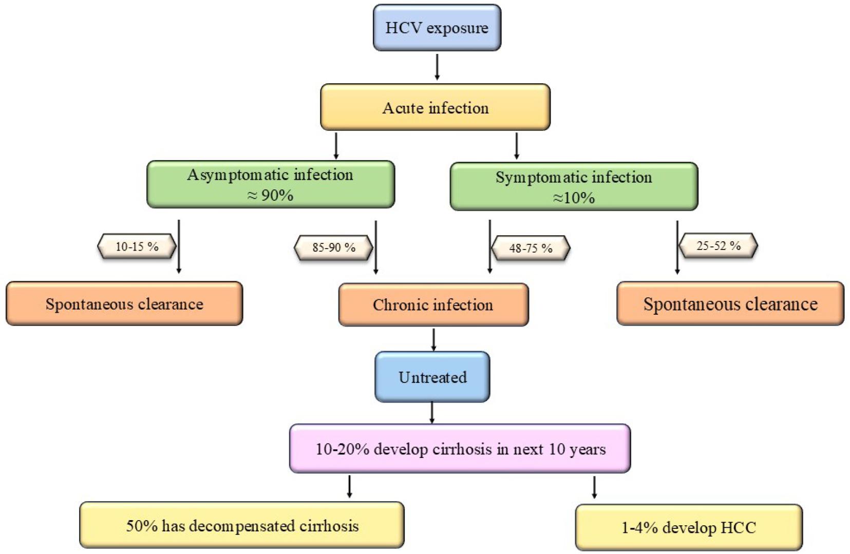
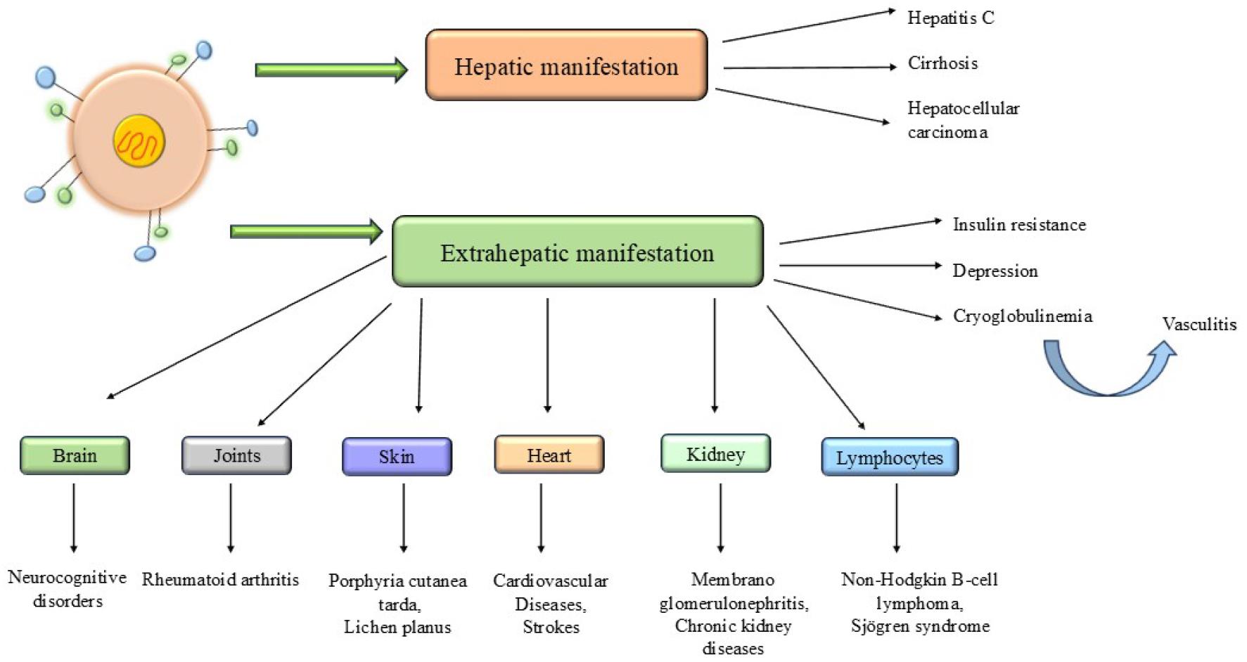
About 90% of acute infection cases are asymptomatic, 10-15% of which are resolved through opsonization. However, most cases progress to chronic HCV infections in the form of liver cirrhosis, HCC, and end-stage liver disease (ESLD) (50) (Fig. 4). HCV has a natural history of hepatotropism and lymphoproliferation (Fig. 5). The hepatotropism accounts for intra-hepatic manifestations like chronic hepatitis C, cirrhosis, and HCC. The lymphotropism is expressed as cryoglobulinemic vasculitis, which is manifested at the neurological, renal, rheumatological, and cutaneous levels (51, 52). The chronic antigen-independent elevation of B cells can even progress to non-Hodgkin B-cell lymphoma (53). In total, hepatic and extrahepatic clinical manifestations account for 10-fold higher deaths in virus-infected patients than in non-infected ones. It suggests the viral infection to be systemic disease and is fatal due to its impact on the immune system.
ORIGIN, PREVALENCE, AND EPIDEMIOLOGY OF HCV
The HCV was originally identified as an infectious agent causing blood transfusion-associated non-A, non-B hepatitis, and was confirmed by studies in chimpanzees. Similar studies led to the discovery of GBV-A, GBV-B, and GBV-C, or hepatitis G virus (HGV), as distant relatives of HCV in New World monkeys. Molecular phylogenetic analysis revealed GBV-B as the closest to HCV. The remaining were classified under a new genus, Pegivirus, within the Flaviviridae family (54). The discovery of a novel guereza hepacivirus (GHV) in colobus monkeys, the first-ever non-human primate hepacivirus, has significantly expanded the host range to Old World monkeys. Although quite divergent from HCV, GHV shares homology with GBV-B, rodent and bat hepaciviruses (55).
Molecular evolution and phylogeny of the virus have wider implications in viral taxonomy, epidemiology, and disease control, leading to a better understanding of infection diagnosis, regimen scheduling, and vaccine candidate design.
Origin of HCV genotypes
The primary basis of the origin of HCV relies on endemicity, recent epidemic transmission, distinct levels of genetic heterogeneity, and the persistent nature of the extant viral strains with variable natural history. A single ancient lineage of the HCV origin has long been constrained due to the lack of evidence of HCV-like infection in non-human primates. However, the occurrence of hepaciviruses in mammals such as bats, wild rodents, horses, and dogs has strongly supported the zoonotic origin theory of HCV. The hepaciviruses isolated from domestic horses and dogs are phylogenetically closest to HCV. Bats and wild rodents are likely the most ancient sources and reservoirs of this lineage of the Flaviviridae family, given their high mobility and global distribution, as well as the remarkable abundance and diversity of hepaciviruses members found in these mammals. Another line of debate lies in the primate versus non-primate source of HCV. The arguments in favor of primate origin lie in the high endemic diversity of distinct genotypes restricted to the areas particularly common for humans, apes, and Old-World monkeys. There is limited transmissibility amongst humans, only through parenteral routes, and a lack of host adaptation as reflected by its severe immune-stimulated liver pathology. However, the absence of any known primate source counters otherwise. Convincingly, evidence supports a non-primate zoonotic transfer event apparently due to a wider host range attributed to its high tolerance to cross-species barriers (56).
The second hypothesis suggests a more recent origin of HCV as an exclusive human pathogen, assuming that each hepacivirus has co-evolved with a specific host species. Throughout evolution, HCV might always have infected mankind; its diversity, however, is a result of hundreds to thousands of years of endemic circulation in specific genotype-dominated geographical areas. Use of host factors for better survivability and enhanced longevity supports the co-adaptation theory. The classic example of such co-evolution theories is the critical dependence of viral RNA on host liver-specific miRNA-122 to complete its replication. The acquired persistence and increased prevalence of permitted transmission routes, as likely, have led to global distribution of HCV (56). Perhaps, HCV has an independent evolutionary lineage since its uncertain origin.
Global prevalence, distribution, and epidemiology of HCV genotypes
The phylogenetic and epidemiological study of worldwide prevalent HCV genotypes has led to certain fundamental rules governing the HCV dissemination and evolution. Substantial genetic variability possessed by the endemic viral strains has been associated with a long course of evolution, whereas the globally represented genotypes with comparatively lower genetic polymorphism and higher rate of transmission, reflect a more recent and shorter course.
Several meta-analyses of the global HCV epidemiology in developed and developing countries presented the viraemic prevalence and genotype distribution (Fig. 6 and Table 2) (57, 58, 59).
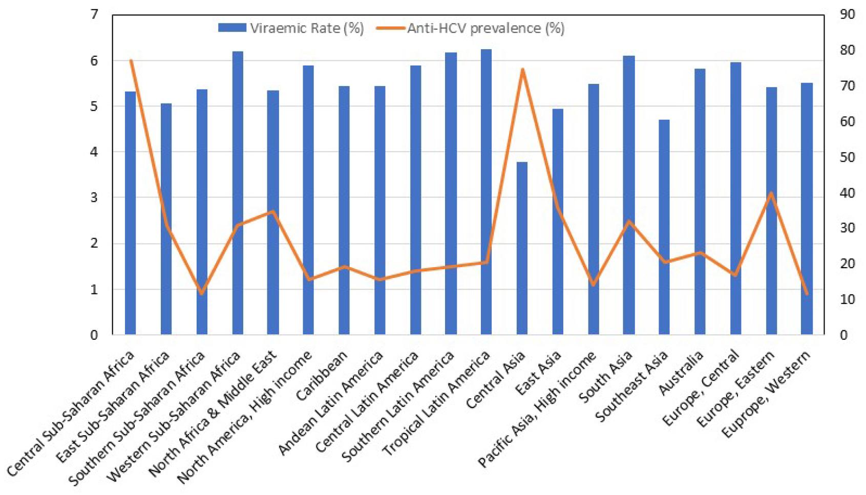
Table 2.
Viremic prevalence and genotype distribution in the countries stratified according to the UN report World Economic Situation and Prospects 2020 (adapted from (60))
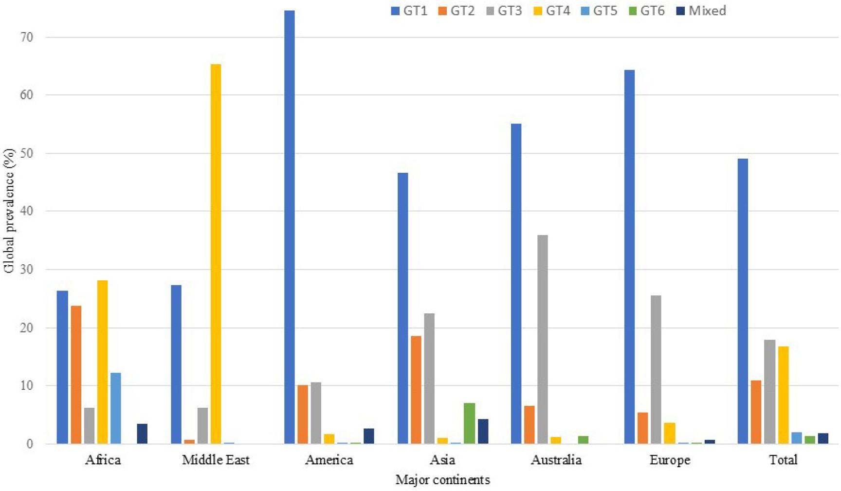
The geographical distribution of HCV genotypes is complex (Fig. 7). Petruzziello et al. (60) suggested the global prevalence to be ~2.5%. Central Africa and Central Asia have been estimated to have a higher prevalence (3.5%) than South Africa, Andean and Central Latin America, North America, Central and Western Europe, and Pacific Asia (<1.5%). Eastern Europe, Australia, Latin America, Caribbean countries, Africa, the Middle-East, South, East, and Southeast Asia have 1.5-3.5% prevalence. The data suggest that HCV GT1 is most prevalent, accounting for 49.1% of global HCV-related infections in adults. It is followed by GT3 (17.9%), GT4 (16.8%), GT2 (11.0%), GT5 (2.0%), and GT6 (1.4%). Mixed or undefined genotypes make up 1.8% of HCV infections. However, differences regarding HCV infection prevalence are recorded at the local, country, and regional levels. The major HCV genotype in Europe, North America, Latin America, and the Caribbean islands is GT1 (64.4%, 66.3%,74.3%, and 83.0%). In the Middle East, North Africa, and Egypt, GT1 (48.3%) and GT4 (32.3%) are the major HCV genotypes. GT1(46.6%) and GT3(22.4%) are the most common genotypes in Asia. GT3 is a prevalent genotype in India (54.4%) and Pakistan (79.0%) (60).
Molecular evolution of HCV genotypes
All the global HCV isolates reported to date have been hierarchically classified into eight major genotypes and more than 90 subtypes. Recently, Mbisa et al. (61) have reported one full-length genome of a novel HCV genotype that fulfils the mandate of ICTV for a new genotype confirmation. Pairwise genetic distance comparison of this variant shows greater resemblance to GT6. However, it forms an equidistant separate clade between GT6 and GT8 in the phylogenetic tree. Also, the same study enlisted GT8b and GT8c subtypes from Indian origin. A genome-wide sequence variability of ~30% and ~15% differentiates between genotypes and subtypes, respectively. Phylogenetic discreteness is the most peculiar feature of genotypes, such that each genotype is represented as an individual clade in the phylogenetic tree. Whereas the subtypes are distinguished by their distribution pattern and high-risk target groups, highlighting their epidemiological importance (62).
The relative evolutionary age of HCV genotypes has suggested GT2 as the oldest one, followed by GT3, GT5, and GT6, whereas GT1 and GT4 branched most recently. A correlation of this order of emergence, when mapped to the genotype biasness, reflected in the duration of treatment and magnitude of clinical response to PEG-IFN/RBV-based therapies. It revealed that older genotypes (GT2, 3, 5, and 6) are more receptive to the corresponding treatment and thus, easier to eradicate. GT2, the oldest genotype, showed SVR maxima, which declined hierarchically in order of GT3>GT4>GT1. It confirms that host immunity is an important factor that has shaped the evolutionary course of HCV, given that humans are the only known natural host of HCV (63).
Point mutation and recombination have played a significant role in the molecular evolution of HCV. Point mutations are the obvious consequence of erroneous replication by HCV RdRp, which lacks the 3′→5' exonuclease activity. Absence of proof-reading leads to higher mutation accumulation rates (10-3 to 10-5 nucleotides/site) during (-)ssRNA-directed (+)ssRNA synthesis. In vitro kinetic studies of NS5B revealed that U:G and G:U mismatches are the most favored and major source of genomic substitutions due to the inherent bias of HCV RdRp. In vivo mutation rates are much lower (2.5×10⁻⁵) largely due to the natural replication and selection pattern. Also, in absence of secondary infections, replication proceeds through multiple replication complexes simultaneously within the infected cell. This means that some of the progeny RNAs can serve as templates indicating the geometric pattern of mutation accumulation. On the other hand, natural selection favours fitter individuals, thereby limiting the otherwise possible mutation spectrum. Lethal mutations can provide near-accurate results only if the nonsense mutations are not complemented by the functional genomes. Notably, the HCV genome has invariable to moderate and high variability regions (HVR1/2) in the E2 regions, unlike the in-vitro replicon systems. It suggests a random mutation pattern throughout the genome. Thus, natural selection has a major role in shaping the overall pattern of mutations across the viral genome (62).
The phenomenon of genetic recombination by the copy-choice replication mechanism is widespread in RNA viruses, but is debatable in the context of HCV due to rare incidences of mixed or superinfections (62). However, a natural inter- genotypic recombinant of HCV was reported and designated as RF12k/1b (64). Since then, nine natural recombinants have been reported (65). Sequence analysis of RF2k/1b revealed NS2/NS3 junction as a recombination hotspot around which template switching by RdRp is maximum. The proposed mechanisms of recombination in HCV are either replicative or endoribonuclease-mediated non-replicative (66). The underestimation of recombination frequency in HCV might be due to the actual low intrinsic recombination rates or the limitations of genotype-targeted diagnostic methods assessed through several in vitro analytical approaches. Such methods have positively indicated a high recombination potential for HCV RdRp, more likely towards highly similar genomes. However, this might not produce the fittest variants with enough viability and/or efficient transmission rates. Thus, despite the high in vitro recombination frequencies, formation of natural recombinants seems to be biologically constricted. An effective co-infection with two equipotent genotypes, crossing over to produce competent circulating recombinant forms (CRFs) coding the complete spectrum of viral proteins, seems critical and rate-limiting. Advanced genotyping assays targeted against multiple pan-genomic sites will be critical in the diagnosis of emerging CRFs.
The concept of quasispecies underlies the foundation of the genetic heterogeneity of HCV. It is defined as a viral subpopulation circulating as a dynamic pool of closely related genetic variants of a master variant infecting the host. This within-host viral diversity is a result of high mutation rates and higher recombination frequencies among these genetically related mutants. Intra-quasispecies recombination might not result in radically distinct genomes, which alongside superinfection exclusion, reasonably explains the sporadic occurrence of CRFs. RF2k/1b being the only intergenotypic recombinant that spread epidemiologically. The quasispecies refers to a complex evolutionary process operating at the host level that assumes genetically variable viral population and, is subject to selection where an interplay of complementation/ suppression maintains an equilibrium between suboptimal variants and the fitter individuals. Though experimental evidence supports the quasispecies nature of the HCV population, many believe that the observed features of the HCV population are a result of group selection in the classical population genetics model.
The HCV evolution is a consequence of viral heterogeneity arising within-host and inter-host levels. It operates under distinct selection pressures and is a function of bottleneck events, which transiently curb the ever-mutating viral population. In a host, the multiplicity of infection (MOI) factor determines the infection course and hence, viral evolution. Generally, high MOI results in enhanced template switching due to the presence of more than one genome template for viral RdRp, leading to greater recombination frequency. Moreover, it confers polyploidy, affecting the phenotypic features of the virus. Thus, MOI can be the primary adaptive strategy opted by the virus to establish productive infection. Post-infection replication rates are critical to HCV evolution since high replication rates facilitate rapid infectivity and greater diversity. This may happen at the cost of higher elimination risks because of vigorous immune responses. On the contrary, lower replication rates ensure prolonged existence, better fitness, and increased rates of transfection. Intra-host heterogeneity of viral quasispecies is subject to further diversification due to its compartmentalization during the spread of infection to extrahepatic sites. These segregated virus populations undergo independent replication to produce distinct quasispecies sub-populations. Interestingly, the quasispecies samples from various tissues revealed that the blood quasispecies has higher infectivity variants, making it the actual transmission-potent population. Thus, the intra-host population steers the evolution at the epidemiological level. Whole-genome sequencing postulated a constant rate of evolution across the genomes, with greater variability observed in E1/E2 coding regions within a single host. However, a sudden decline in this heterogeneity upon the change of host ensures a better scope of adaptability in the new host. Moreover, the HCV subtypes differ in rates of evolution, as GT1a is reported to evolve faster than subtype GT1b. Thus, the intergenic rate of evolution within a genome are mutually dependent and varies majorly as a function of selection pressure that has significant implications at the epidemiological level. In nature, genes under extreme immunogenic selection pressure tend to evolve at a greater rate as compared to those with constrained selection pressure. It is well exemplified by the hypervariable regions encoding for HCV surface antigens (E1/E2 glycoproteins) and the relatively conserved regions encoding the replication proteins (NS3, NS5B), respectively. Host immunity and the anti-viral drugs are two critical selection forces that drives HCV evolution at the host level and defines the success rate of treatment regimes. Tolerance to such selection pressures is usually manifested as genome-wide nucleotide substitutions (RASs). Majority of these mutations are spontaneous and lead to natural genetic diversity in viruses at individual, population and global levels. The natural occurrence of RAS-variants as minor group in within-the-host viral quasispecies is one of the adaptive strategies that viruses employ to escape host immunity as well as drug selection pressure. Despite of the induced therapeutic pressure, HCV replicase is biased to mutate genomic regions that can provide greater resistance at the minimum fitness cost. The non-structural proteins such as NS3/NS4A protease, NS5A replication complex and NS5B polymerase are particularly the prominent DAA-targets and are prime hotspots of RAS mutations. Such SNPs weakens the enzymatic functions of the protein rendering them unfit for selection. However, the erroneous high replication rates inherent to HCV polymerase enable the virus to trade-off between drug evasion and selection fitness by incorporating genome-wide compensatory mutations that improves fitness while maintaining drug resistivity. In general, the drug-resistant mutations are well tolerated in NS5A replication complex and hence these variants are more likely to have long term evolutionary persistence with greater risk of drug tolerance and treatment failures. On contrary, since the fitness cost is extremely high for NS3/NS4A proteases and NS5B polymerase, these RASs undergo purifying selection or can revert back to wild-type genotype under consistent drug pressure. Data for global prevalence of RAS-associated variants (RAVs) showed geographical as well as genotypic biasness. RAVs represented a significant portion of all genotype population with different geographical frequencies. Amongst genotypes, highest frequency was recorded for GT6 (99%) followed by GT2> GT4>GT1a>GT3>GT1b with 87.9%, 85.5%, 56%, 50% and 34.3% prevalence. Across continents, Asia was recorded with highest frequency of 74.1% and Europe at minimum with 51.4% (67). Global discrepancies in RAVs distribution are the apparent result of distinct genotypic composition of viral populations found in these locations along with the demographic factors, major transmission routes, prevalence of high-risk behaviors among infected population and the extent of accessibility to diagnostic and therapeutic regimes. The HCV evolve extraordinarily under DAA selection pressure. Existence of HCV as a quasispecies in the host is critical to the clinical outcomes of therapeutic regimes. Administration of DAAs has a bottleneck effect leading to clearance of the major susceptible viral subpopulation. This, however, also confers a selective advantage to the pre-existing small viral population with baseline RAS that counter the drug susceptibility. Subsequently, the rapid and erroneous replication of HCV drive directional evolution of the existing mutants into a dominant therapy-resistant population. Although this resistivity comes at cost of compromised replication fitness, it is compensated over the time by the incorporation of substitutions in surrounding genomic regions to restore the functional fitness of proteins and enhance the viral vitality. The DAA-mediated RAS selection pressure pose a serious challenge to long-term viral elimination goals. The current pan-genotypic DAA therapies co-administered with anti-viral drugs target multiple genotypes, thus, render the accumulation of precise mutations in viral population simultaneously against multi-drug selection pressure. Current DAA therapies have simpler dosing schedule, minimal side effects, shorter course of treatment and highest SVR of all the anti-viral therapies. The most recommended co-formulations with non-overlapping RAS-targets includes glecaprevir/pibrentasvir and sofosbuvir/velpatasvir. Their broad therapeutic window has simplified the treatment landscape (68).
CLINICAL RELEVANCE OF HCV GENOTYPES
From IFN-α monotherapy, through combined IFN-α/ribavirin therapies, to the development of current DAA strategies with high SVR rates has made hepatitis C a curable disease provided timely diagnosis is made. There is a certain relation between HCV genotype, its natural history, disease prognosis, and the type of response generated against administered antiviral therapies. IFN-based therapies were found to be more effective, with higher SVR rates and shorter treatment duration, in GT2 and GT3 compared to GT1 and GT4 (69). Both GT1 and GT3 are ranked highest in terms of pathogenicity, being associated with the most severe cases of post-cure liver disease progression. Host factors such as age, gender, liver histology, and infection duration also influence disease severity and treatment effectiveness.
A plausible association between HCV genotype and infection outcome is one of the most intensively studied areas, aiming to establish clinical correlation with HCV genotypes. These include progression of liver fibrosis, development of cirrhosis, association of extrahepatic diseases with hepatitis C, recurrence after post-liver transplantation, and the impact of HCV genotypes on co-morbid diseases like HIV.
GT1 has been linked to aggressive liver fibrosis and slower antiviral responses to IFN/RBV-based therapies, though it responds effectively to DAAs (70). GT1-infected individuals with specific IL28B gene polymorphism respond more effectively to PEG/IFN-α/RBV therapies- an example highlighting the significance of host-virus interactions in determining treatment efficacy. Bruno et al. (71) demonstrated that patients infected with GT1b are at a relatively higher risk of developing HCC. Multivariate statistical analysis confirmed that GT1b is independently correlated with neoplastic transformation and associated with older age and cirrhosis (72). However, the risk of HCC in GT1b-infected patients may be independent of cirrhosis, possibly due to the oncogenic potential of HCV (73). Further studies support the influence of HCV genotypes on the clinical course of infection. For instance, recurrence of hepatitis C in liver allografted patients showed that GT1b infection led to more aggressive liver-related pathologies than other genotypes, indicating that the viral genotype is a critical prognostic factor in transplant settings (74). The mechanisms underlying the greater virulence of GT1 remain unclear, but may involve enhanced immune escape through an additional hypervariable region in the E2 protein or distinct regulatory mechanisms.
Data from a cohort study dominated by GT3 infections linked this genotype to more severe liver steatosis and disturbances in lipid and sugar metabolism, resulting in higher incidences of overall liver disease progression (OLDP). OLDP encompasses accelerated liver fibrosis, increased cirrhosis, HCC, and liver-related mortality (75). While proponents attribute this aggression to genotypic-specific features, critics argue it is associated with metabolic profile and socio-behavioural patterns of the population, given the widespread occurrence of GT3 among intravenous drug users (IDUs) and incarcerated individuals. This is supported by the relatively low independent statistical weight of genotype in multivariate models correlating infection and substance use in defined age groups. Moreover, GT3 has been linked to aggressive liver fibrosis progression due to the viral interference with host lipid metabolism, causing severe hepatic steatosis even in non-obese, non-diabetic patients. This worsens prognosis and necessitates longer treatment courses (76). Nonetheless, genotype- specific hepatic complications may interact with other virulence-influencing factors, such as the host population and environment. Global pan-genotypic studies suggest that the severity of liver diseases is likely independent of causative genotypes.
After GT1 and GT3, GT2 subtypes show the highest global prevalence, fastest viral clearance, and slower progression to chronicity. Other genotypes remain largely endemic. The disease course is a cumulative outcome of viral and host genomics. For example, a study of GT1-infected patients suggested that IL28B (IFN-λ3) gene polymorphism is an indicator of IFN therapy success, with unfavourable C/T and T/T genotypes responding poorly, yielding low SVR and higher risk of treatment failure, compared with favourable C/C genotypes (rs12979860) (77). Ubiquitin-specific protease 18 (USP18), an interferon-stimulated host gene that negatively regulates IFN-related antiviral defence, has been shown to potentiate antiviral activity upon silencing (78).
The main controversies revolve around the confounding association of HCV genotypes with the distinct routes of disease development, clinical behaviours, and post-treatment risk of OLDP. While genotype bias is central to these variables, it is not the sole controlling factor. Genetic variability is crucial to the immunologic escape routes adopted by the virus, which in turn determines the course and severity of infection. Host genetic factors such as polymorphisms in IL28, HLA, and IFNL4 shape HCV evolution and are key determinants of treatment response (79). Thus, HCV disease progression represents a complex interplay of viral and host genetics, further influenced by the environmental variables. Arguments in support of genotype-driven progression suggest that epigenetic and structural genetic modulation induced during chronic infection determines the probability of developing post-cure liver diseases. Conversely, arguments against emphasize that baseline liver injuries (Fibrosis, F3/F4) present at the time of treatment are the primary drivers of late-stage liver diseases, rather than genotype-dependent oncogenic footprint. Nonetheless, the paradigm shift from IFN to DAA therapies has drastically reduced the pathological impact of HCV genotypes due to their pan-genotypic efficacy.
CURRENT STATUS OF AVAILABLE TREATMENTS
The HCV treatment started with IFN-α monotherapy against an unidentified post-transfusion-associated non-A/non-B hepatitis in 1986. Although it showed several side effects for chronic infections, lower efficacy, and recurrence of infection upon cessation of medication, sustained high SVR (~98%) popularized it as the primary choice for acute infection treatment (80).
Major chronic infection treatment involved ribavirin, a broad-spectrum antiviral nucleoside analog. It decreased the alanine aminotransferase (ALT) levels, suggesting an effective oral treatment. However, the subsequent modulations in host nucleotide metabolism and low SVR rendered it unpopular. Later on, administration of IFN-α with ribavirin led to a remarkably higher SVR rate of 40-50% for GT1 and up to 80% for GT2, decreased ALT levels with improved hepatic histology in comparison to ribavirin or INF-α monotherapy. This proved to be a breakthrough in the treatment of chronic infections (81). PEGylated interferon combined with ribavirin, with an impressive 52% SVR, was another milestone in HCV treatment (82). However, higher mutation rates in HCV results in quasi-species viral variants that can deceive immune responses. Thus, INF-α monotherapy is highly vulnerable against genetically variable HCV. Deeper insights into the HCV life cycle suggested the use of protease inhibitors against non-structural viral proteins, marking the new era of DAAs which revolutionized the HCV treatment with significantly improved SVR rates as compared to prior therapies. Telaprevir and boceprevir (serine protease inhibitors targeting NS3/4A) were the first-generation approved DAA regimens (83, 84, 85) that marked a significant milestone (Table 3). Combinations of these DAAs with PEG-IFN and ribavirin, boosted response rates in patients with GT1 infection. Second-generation DAAs, like ledipasvir and sofosbuvir, offered IFN-free regimens with enhanced efficacy and safety profiles. Since then, several DAAs formulated against the viral genome (Table 4) have revolutionized the HCV treatment landscape with improved tolerance, high response rate (>95%) (86, 87) and simplified the management of HCV treatment. It led to a significant improvement in long term health-related quality of life. Development of pan-genotypic DAAs designed to target multiple genotypes is a remarkable advancement in HCV therapy. Pan-genotypic DAAs like sofosbuvir-velpatasvir, glecaprevir-pibrentasvir, and other regimens simplified the treatment perspective that offer reduced genotype pretreatment testing, shorter treatment durations and fewer side effects. The SVR rates and safety profiles of pan-genotypic DAAs against HCV are excellent. These DAAs achieved higher SVR rates across all genotypes, and improved tolerability even in chronic HCV infections. Another pan genotypic DAA combination of sofosbuvir/velpatasvir/voxilaprevir achieved an SVR of 88.6% and was shown to be an effective rescue therapy for failures to sofosbuvir/velpatasvir or glecaprevir/pibrentasvir treatment (88). Despite the overall high success rate of DAA therapy, clinical observations show that some patients respond poorly to DAAs or experience virological relapse. Such cases are mainly GT3 or have coexisting cirrhosis, and their poor SVR may be associated with resistance-related substitution mutations in HCV genome, especially in the NS5A region (89).
Table 3.
DAAs of different generations for HCV therapy (Adapted from (68))
Table 4.
Comparison of HCV therapy with pan-genotypic DAAs (68)
Moreover, the existence of quasispecies further expands the scope of recombination-driven genetic variability. The elusive mechanism of T-cell action against viral persistence, lack of effective cell culture models, absence of conventional animal model due to a narrow host range, and ethical concerns regarding animal use, particularly chimpanzees for broader T-cell response, and CD4+ and CD8+ memory cells to generate a sustained response (90) are major challenges toward the development of a promising vaccine. Due to the potential risks associated with attenuated live-virus vaccines, highly efficacious and safer vaccine candidates based on viral genes and/or proteins have been investigated (91). While HCV structural proteins (E1, E2, and core) generate neutralizing antibody (nAb) responses, non-structural proteins have been shown to induce stronger CD4+ and CD8+ T-cell responses. CD8+ T-cell induction also assists in the production of cross-neutralizing antibodies. Nevertheless, HCV envelope glycoproteins remain the most extensively studied and popular subunit vaccine candidates, tested singly or in combinations (E1, E1/E2, and/or E1/E2-Core) (38). Clinical trials using mouse models, non-human primates, and human have been conducted to assess efficacy. Despite encouraging initial results, none have advanced to phase III human trials to date (https://clinicaltrials.gov/). Further, the mRNA vaccines hold greater promise due to their ability to elicit both humoral and cellular immune responses. They combine the advantages of subunit vaccines (safety and precise composition) with those of live attenuated vaccines (internal antigen presentation and T-cell activation). Their direct interaction with pattern recognition receptors (PRRs) results in robust innate immune activation. Moreover, the avoidance of virulence reversion and insertional mutagenesis makes mRNA vaccines a safer choice. Recent approaches to vaccine design include recombinant protein vaccines, E1/E2-based subunit vaccines, synthetic peptide vaccines, dendritic cell-based vaccines, cell culture-based vaccines, virus-like particles (VLPs), DNA vaccines, and recombinant virus vaccines. Most of these, however, are in the preclinical study phase. Meta-analysis of the efficacy of potent anti-HCV vaccines in chimpanzees suggests that whole or a part of the E1 and/or E2 glycoprotein region may elicit cellular and humoral immune responses (92). An ideal vaccine should elicit multivalent immune responses to counter the inherent protein variability across all HCV genotypes and induce vigorous VnAb responses against the E1/E2 glycoproteins (93, 94). Another meta-analysis in chimpanzees also indicated that the use of complete or partial E1/E2 glycoprotein resulted in a better immune response (92). The prime attraction for humoral immune response against HCV is the use of antigenic epitopes from whole or part of core, E1, and E2 proteins (91). In recent years, various potent vaccines have been tested in primates and mice, though only a few have qualified for human trials (95). As it is, major limitations include limited cellular and humoral response, less effective adjuvants, and low delivery of immunologically important viral epitopes (96). The HCV carrier percentage in chimpanzees immunized with E1/E2 was much less than that of chimpanzees used as controls (97), with elevated levels of VnAbs titres against GT1a, which in vitro neutralized both HCV cell culture (HCVcc) and HCV pseudoparticles (HCVpp) consisting of E1/E2 envelope proteins from different genotypes (98). Adeno-virus, glycoprotein-based vaccines, primed and boosted with adjuvants, were able to elicit stronger T-cell responses and a higher amount of VnAbs in rodents (94). Thus, it can be inferred that the genetic hypervariability of the virus can be better targeted by the use of multi-epitopic DNA vaccines and even better with an adjuvant-coupled peptide vaccine (99). However, challenges such as low translational efficiency, poor stability, and delivery system limitations remain significant (100). The lack of efficient delivery systems for many clinically evaluated mRNA vaccines underscores the urgent need for improvement in vaccine delivery technologies. Current vaccine strategies, their viral protein targets, specific immune responses, and clinical trial stages are summarized in Table 5.
Table 5.
Prophylactic and therapeutic HCV vaccines in human clinical trials (38)
The current global scenario of HCV epidemiology emphasizes the necessity of a potent pan-genotypic anti-HCV vaccine in curbing the early infection rates, while advancement in DAA therapies with novel therapeutic target(s) to overcome RAS effects. The emergence of CRFs has further challenged the current diagnostics and therapeutics, thereby raising the demand for a novel and holistic approach, including recombinant mRNA-based vaccines, which are not only capable of inducing a robust T-cell response but also provide durable humoral immunity.
SIGNIFICANCE OF GENOTYPE SURVEILLANCE IN CONTEXT OF GLOBAL HCV ELIMINATION EFFORTS
Advent of pan-genotypic DAAs have revolutionized the anti-HCV therapy and has become the general first-line of medication to all those patients who qualify under simplified treatment categories without any cirrhosis compensation and history of treatment failures or re-infection. This line of treatment has transformed the clinical procedures to simple ‘test-and-treat’ pathways bypassing the barrier of tedious genotyping to access HCV care. Despite the development of highly effective antiviral treatments, the WHO’s target of eliminating HCV by 2030 faces major hurdles of reinfection among high-risk groups, large gaps in diagnosis, limited healthcare accessibilities and persistent challenges in reaching vulnerable populations.
In line with the HCV elimination goal, addressing these systemic issues are of utmost importance. Genotype surveillance is important for tracking the viral transmission network at macro-levels to gauge the shifts in genotype-spectrum and associated risk parameters across the high-risk groups like people who use drugs (PWUD), prisoners, migrants, marginalised communities, individuals exposed to unsafe medical practices, and those with co-infected with HIV/HBV. Such communities are disproportionately affected and remain vulnerable to reinfection after successful treatment and undermine the overall progress. Globally, over 70% of HCV-infected individuals remain undiagnosed, largely due to asymptomatic early stages. This gap worsens due to limited screening programs; lack of awareness and stigma prevent timely detection. In such instances, genotype testing is paramount to distinguish between cases of new infections, relapse due to pharmaceutical failure, and re-infection due to high-risk exposure as it help healthcare authorities to streamline their approach as per the real cause. Tracking real-time dynamics of viral circulation amongst smaller communities, endemic to specific genotypes render public health centres to deploy targeted interventions at micro-levels against the specific viral population. The tailored intervention such as expansion of needle-exchange programs, opioid substitution treatment, and safe medical practices, scaling up affordable and rapid diagnosis and integration of HCV screening into routine healthcare are essential. Moreover, it also important for prognostic stratification for better prediction of the post-cure pathological risks, sustainable allocation of resources and effective management of long-term disease burden. The existing disparity in the geographical meta-data of viral sequencing creates a blind spot masking the actual instances of viral infection and circulation status in areas with least or no sampling. Also, underrepresentation of rare and endemic strains in the global genomic databases further causes a prejudice in diagnostic and treatment strategies. Thus, an active genotype-based surveillance system for uniform tracking of all HCV variants circulating globally is key to achieve the global elimination targets.
CONCLUSION
HCV is a genetically diverse and epidemiologically complex human pathogen exemplifying the interplay of viral evolution and host adaptation. Its enigmatic evolutionary origin continues to challenge the virologists. The global distribution of HCV genotypes highlights both ancient and endemic circulation and recent epidemic spread, with GT1 and GT3 dominating worldwide prevalence. Molecular evolution driven by high mutation rates, quasispecies dynamics, and sporadic recombination events has structured its adaptability and therapeutic responses. Clinically, genotype-specific differences influence disease progression, risk of HCC, and treatment efficacy, though the advent of DAAs has largely overcome genotype bias. Despite remarkable therapeutic advances, vaccine development remains hindered by HCV’s extraordinary heterogeneity and immune evasion strategies. Current research emphasizes multi-epitopic, pan-genomic vaccine approaches, including DNA- and mRNA-based scaffolds to achieve protective humoral and cellular immunity and subsequent eradication of HCV.