


INTRODUCTION
Coxsackievirus B3 (CVB3), a positive-sense single-stranded RNA virus of approximately 7.4 kb belonging to the Picornaviridae family, is the primary causative agent of severe viral myocarditis in children and young adults (1, 2, 3). CVB3 initiates infection by binding to the coxsackievirus and adenovirus receptor (CAR) and decay-accelerating factor (DAF/CD55) on the host cell surface, facilitating viral entry into cardiomyocytes (1, 4, 5). Upon cellular entry, the virus utilizes its encoded 2A and 3C proteases to synthesize viral proteins; simultaneously, these proteases directly induce structural disruption and functional impairment in cardiomyocytes by cleaving host cytoskeletal proteins and translation initiation factors (6, 7, 8).
Following this primary virus-mediated cytotoxicity, the infection triggers a massive infiltration of immune cells, including macrophages and T cells, culminating in a secondary hyperimmune response. If the ensuing extensive inflammation and myocardial necrosis are not properly resolved, ventricular remodeling accelerates, leading to irreversible dilated cardiomyopathy and fatal heart failure (9, 10, 11). Currently, there are no approved targeted therapies in clinical practice that directly inhibit CVB3 replication. Therefore, an urgent need exists for novel therapeutic strategies capable of simultaneously suppressing viral replication and mitigating the host’s pathological immune response.
Recently, emerging evidence in the pathogenesis of viral myocarditis has revealed that CVB3-induced cellular damage is primarily amplified by pyroptosis, a highly inflammatory form of programmed cell death rather than simple apoptosis or necrosis (12, 13). During infection, the intracellular accumulation of viral RNA and concurrent viral-induced cellular stress stimulate the pattern recognition receptor (PRR) NLRP3. Upon activation, NLRP3 associates with the adaptor protein ASC to recruit pro-caspase-1, culminating in the assembly of the multiprotein NLRP3 inflammasome complex (14). Inflammasome assembly triggers the autoproteolytic cleavage and subsequent activation of caspase-1, which then executes two critical downstream functions. First, it processes the pro-inflammatory cytokine precursors pro-IL-1β and pro-IL-18 into their biologically active, mature forms. Second, active caspase-1 cleaves gasdermin D (GSDMD), the central effector protein of pyroptosis, separating its autoinhibitory C-terminal domain from the pore-forming N-terminal domain (GSDMD-N) (15, 16). The liberated GSDMD-N translocates to the plasma membrane, where it oligomerizes to form pores with a diameter of 10–15 nm. These pores facilitate the explosive extracellular release of mature IL-1β, IL-18, and various damage- associated molecular patterns (DAMPs), ultimately culminating in osmotic imbalance, plasma membrane rupture, and the completion of pyroptotic cell death (12, 16). Consequently, therapeutically targeting this CVB3-induced pyroptotic pathway represents a highly promising strategy for attenuating excessive myocardial inflammation and preserving host cell integrity.
Houttuynia cordata, a perennial herbaceous plant belonging to the Saururaceae family, is widely distributed across wetland habitats in East Asia, including Korea, China, and Japan. Historically, it has served as a pivotal medicinal herb in traditional medicine for the treatment of various infectious and inflammatory conditions, such as pneumonia, respiratory infections, and enteritis. Phytochemical profiling has revealed that H. cordata is exceptionally rich in flavonoids and polyphenolic compounds, including quercetin, quercitrin, rutin, afzelin, and hyperoside, as well as essential oils like decanoylacetaldehyde (17, 18). These primary bioactive constituents exert antiviral effects via multifaceted mechanisms, such as interfering with viral attachment and host cell entry and inhibiting critical enzymatic activities required for viral replication. Extensive prior research has demonstrated that H. cordata extracts exhibit broad-spectrum and potent antiviral efficacy against a diverse array of RNA and DNA viruses. These include herpes simplex virus (HSV), influenza virus, severe acute respiratory syndrome coronavirus (SARS-CoV), dengue virus, and enterovirus 71 (EV71), a member of the Enterovirus genus closely related to CVB3 (19, 20, 21, 22). Furthermore, H. cordata is highly regarded for its profound anti-inflammatory and immunomodulatory properties. It has been shown to downregulate the NF-κB and MAPK (mitogen-activated protein kinase) signaling pathways in immune cells, particularly macrophages, thereby attenuating the hypersecretion of pro-inflammatory cytokines and protecting tissues from inflammatory damage. Consequently, H. cordata has emerged as a highly promising natural therapeutic candidate for managing infection-driven inflammatory pathologies (23).
Despite the well-documented antiviral and anti-inflammatory properties of H. cordata, its direct inhibitory efficacy against CVB3 infection and, more specifically, the precise molecular mechanisms underlying its modulation of CVB3-induced pyroptosis remain poorly elucidated. Therefore, the present study was designed to evaluate the antiviral effects of H. cordata extract on CVB3 infection and viral replication in vitro. Furthermore, we aimed to delineate the underlying regulatory mechanisms through which the extract confers cytoprotection, specifically focusing on its capacity to suppress caspase activation and GSDMD-mediated pyroptosis in a CVB3-infected cell model. The findings of this study are expected to provide a robust scientific rationale for the development of safe, natural product-derived therapeutics to effectively manage viral myocarditis and severe inflammatory diseases triggered by CVB3 infection.
MATERIALS AND METHODS
Cell culture and viruses
HeLa cells, a human cervical cancer cell line, were cultured in Dulbecco’s Modified Eagle Medium (DMEM, WELGENE, Korea) supplemented with 5% Fetal Bovine Serum (FBS, WELGENE, Korea) and 1% Penicillin-streptomycin at 37°C under 5% CO₂ conditions. Cells were subcultured at intervals of 4 days and maintained for use in experiments. Coxsackievirus B3 (Woodruff strain, provided by Dr. Eun-Seok Jeon) was used to infect cultured HeLa cells for 18 hours, and the supernatant was obtained to check the virus concentration. A plaque-forming unit (PFU) assay was then performed, and the supernatant was diluted to a concentration of 104 PFU, which was the concentration to be used in the experiment (24).
Preparation of H. cordata extract
H. cordata leaves were washed three times with distilled water to remove impurities, pre-frozen in an ultra-low temperature freezer at -80°C, and then completely dried using a lyophilizer. The dried leaves were ground into a uniform powder using a grinder, and 50 mL of distilled water was added to the powder to perform water extraction at 121°C for 2 hours. The extract was filtered through filter paper and concentrated using a rotary evaporator. The concentrated extract was freeze-dried again to produce the final H. cordata water extract (HCWE) powder. The prepared extract powder was stored at -20°C until use; immediately before the experiment, it was dissolved in cell culture medium or PBS at a concentration of 10 mg/ml, filtered through a 0.22 μm syringe filter, and then used (25).
Toxicity and Antiviral Activity Evaluation of H. cordata Extract
To evaluate the toxicity of the H. cordata extract itself, cells were seeded into a 96-well plate at a concentration of 3 x 10³ cells/well and incubated for 18 hours. Subsequently, the supernatant was removed, and the extract was applied at concentrations ranging from 1000 µg to 0.01 µg/ml. The cells were then incubated at 37°C for 18 hours. Additionally, 7 µl of cck8 (Cell Counting Kit-8) was applied to each well at 30-minute intervals for 2 hours, after which the absorbance was analyzed to confirm the toxicity of the extract.
Western blot analysis
Proteins from HeLa cells treated with the virus and extract were loaded onto 10% or 12% acrylamide gels and subjected to electrophoresis at 80–100 V for approximately 90 minutes. Subsequently, the samples were transferred to a polyvinyl difluoride (PVDF) membrane at 250 mA at room temperature for 2 hours. After transfer, the amount of protein transferred to the membrane was confirmed by Ponceau staining, and non-specific antibody reactions were removed by incubating in 5% non-fat milk blocking buffer for 1 hour. Primary antibodies, such as CVB3 VP1 (Cell Signaling, USA) and GAPDH (Cell Signaling, USA), were incubated at 4°C for 16 hours, followed by the incubation of a secondary antibody targeting Horseradish Peroxidase (HRP). The membrane was treated with ECL solution (Intron Biotech, Korea), and observed using Bio-Rad Chemi-DOC.
Immunofluorescence staining
Immunofluorescence staining was performed to analyze the relationship between cytoviral infection and the induction of pyroptosis. Hela cells were cultured in 12-well plates fitted with cover glass and treated with H. cordata extract and CVB3 virus for 24 hours. Afterward, the HeLa cells were fixed by treating them with 4% paraformaldehyde (PFA) at room temperature for 15 minutes. The fixed cells were washed three times with wash buffer (1X PBS, 0.2% BSA), treated with a blocking solution containing 0.2% Triton X-100 and 1% BSA for 1 hour, and then blocked with 1% BSA solution for 1 hour to prevent non-specific binding. Primary antibody specific to the target proteins (CVB3 VP1, IL-18) was incubated at 4°C for 18 hours. After washing with wash buffer, a secondary antibody conjugated with a fluorescent substance was incubated at room temperature in the dark for 1 hour. Cell nuclei were counterstained with DAPI solution for 10 minutes. Once staining was complete, the cells were fixed onto a slide glass using mounting medium. Images were acquired and analyzed using a fluorescence microscope.
Statistical Analysis
All results are expressed as mean ± SD. Statistical significance was evaluated using the unpaired Student’s t-test for comparisons between two means. Differences between groups were evaluated using one-way ANOVA by Dunnett’s post hoc test for multiple comparisons against the virus-only control group. (GraphPad Prism 8.0 for Windows; GraphPad Software, La Jolla, USA). P values of less than 0.05 were considered statistically significant.
RESULTS
Evaluation of the Cytotoxicity and Antiviral Efficacy of H. cordata Extract
First, the in vitro cytotoxicity of the H. cordata extract on host HeLa cells was evaluated. Following the treatment of HeLa cells with the extract at concentrations ranging from 1000 to 0.01 µg/mL, cell viability assays revealed no significant cytotoxicity or cell death at any of the tested concentrations compared to the untreated control group (Blank) (Fig. 1A). This demonstrates that the H. cordata water extract possesses a highly favorable safety profile in host cells at concentrations up to 1000 µg/mL. The antiviral efficacy of the H. cordata extract against CVB3 infection was investigated. In the virus-only infected group lacking drug treatment, cell viability plummeted to less than 50% of that observed in the normal, uninfected control (Blank). However, therapeutic intervention with the H. cordata extract following CVB3 infection effectively attenuated virus-induced cell death. Notably, cell viability was restored in a striking dose-dependent manner as the concentration of the extract increased (Fig. 1B). The maximal cytoprotective effect was observed at the highest tested concentration of 1000 μg/mL, thereby confirming that the H. cordata extract robustly inhibits CVB3 infection and viral replication.
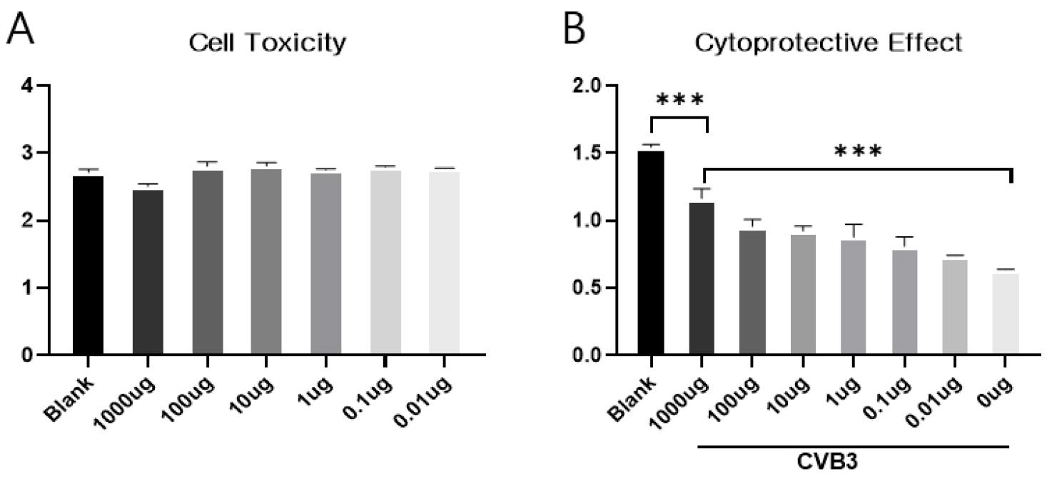
Fig. 1
H. cordata extract exhibits a favorable safety profile and confers dose-dependent cytoprotection against CVB3 infection. (A) HeLa cells treated with H. cordata extract (1000-0.01 µg/mL) for 18 h exhibited no significant cytotoxicity at any concentration compared to the untreated controls, demonstrating a favorable safety profile. (B) CVB3-infected HeLa cells treated with H. cordata extract showed a dose-dependent restoration of cell viability, with maximal protection observed at 1000 µg/mL, whereas virus-only infected cells (0 µg/mL) exhibited <50% viability. Data are presented as the mean ± SD (n = 3). P < 0.001 vs. the indicated groups.
Inhibition of CVB3 RNA replication by H. cordata extract
To elucidate the antiviral mechanism of H. cordata extract at the transcriptional level, strand-specific RT-PCR was performed to differentiate and evaluate the synthesis of CVB3-positive strands (viral genome) and negative strands (replication intermediates) RNA (Fig. 2A). Experimental results showed that while both RNA strands were strongly amplified in the virus-alone infection group, the H. cordata extract treatment group significantly inhibited the synthesis of both RNA strands in a dose-dependent manner. Densitometric quantitative analysis (Fig. 2B) revealed that the group treated with the extract at a concentration of 1000 µg/mL showed a significant decrease in the expression of negative-strand RNA, a key indicator of active viral replication, compared to the infected control group. Consequently, these results suggest that H. cordata extract effectively blocks the intracellular RNA replication mechanism of CVB3.
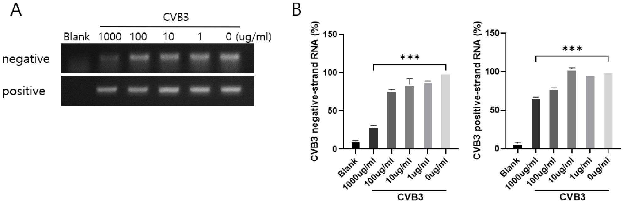
Fig. 2
H. cordata extract inhibits CVB3 viral RNA replication. HeLa cells were mock-infected (Blank), infected with CVB3 alone (0 µg/mL), or infected and treated with H. cordata extract (1000-1 µg/mL). Strand-specific RT-PCR analysis was performed at 18 h post-infection. (A) Representative agarose gel images showing the amplification of positive-strand (genomic) and negative-strand (replicative intermediate) viral RNA. (B) Densitometric quantification of the viral RNA bands. Viral RNA synthesis was robustly elevated in untreated infected cells but dose-dependently suppressed by H. cordata treatment, showing approximately a 40% and 70% reduction in positive- and negative-strand RNA at 1000 µg/mL, respectively. The relative RNA levels are expressed as a percentage relative to the virus-only group (100%). Data are presented as the mean ± SD of three independent experiments. P < 0.001 vs. the virus-only group.
Inhibitory Effects of H. cordata Extract on CVB3 Replication and Host Protein Alterations
We performed Western blot analysis to investigate whether the extract’s cytoprotective benefits come from directly blocking viral replication and modulating downstream pathological signals. The experimental groups consisted of normal cells mock-infected (Blank), cells infected solely with CVB3 (0 μg/mL), and cells infected with CVB3 followed by treatment with various concentrations of the extract (1000-1 μg/mL). As expected, the major viral capsid protein CVB3 VP1 was highly expressed in cells infected with the virus alone. However, treating the cells with the H. cordata extract led to a marked drop in VP1 levels, which decreased further as the extract dose went up. At the maximum dose of 1000 μg/mL, this viral protein became nearly undetectable compared to the untreated infected controls (Fig. 3A). These results suggest that the H. cordata extract directly disrupts CVB3 replication and viral protein production inside the host cells. To hijack the cellular machinery for its own use, CVB3 typically breaks down the host translation factor eIF4G1. We observed a clear drop in intact eIF4G1 in the virus-only group; remarkably, adding 1000 µg/mL of the extract prevented this breakdown, keeping eIF4G1 at the levels seen in the uninfected controls (Blank). We also tracked PARP cleavage—a standard marker for apoptosis—which spiked in the CVB3-infected group due to severe virus-induced cellular damage. Applying the extract at 1000 µg/mL drastically lowered this cleavage, bringing it down past even the baseline mock-infected levels (Fig. 3A, B). In summary, H. cordata extract goes beyond merely keeping infected cells alive to actively disrupt viral action at the molecular level. It can be confirmed that antiviral action occurs by protecting host protein synthesis mechanisms through the inhibition of viral VP1 production and by inhibiting the infection-induced PARP-mediated apoptosis pathway.
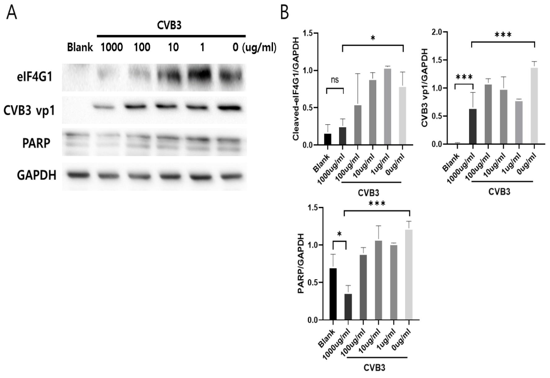
Fig. 3
H. cordata extract inhibits CVB3 replication and protects the host translational machinery from virus-induced degradation. HeLa cells were mock-infected (Blank), infected with CVB3 alone (0 µg/mL), or infected and treated with H. cordata extract (1000-1 µg/mL). Western blot analysis was performed at 18 h post-infection. (A) Representative Western blot images showing the expression levels of eIF4G1, CVB3 VP1, and PARP. GAPDH served as the loading control. (B) Densitometric quantification of the Western blot bands relative to GAPDH. The CVB3 VP1 capsid protein was robustly expressed in untreated infected cells but was dose-dependently suppressed by H. cordata treatment, with near-complete abolition at 1000 µg/mL. Furthermore, the virus-induced degradation of eIF4G1 (eukaryotic translation initiation factor 4G1) and the cleavage of PARP (an apoptosis marker) were substantially attenuated by the extract in a dose-dependent manner. Data are presented as the mean ± SD of three independent experiments. ns, not significant; *P < 0.05, ***P < 0.001 vs. the indicated groups.
Inhibitory Effects of H. cordata Extract on the CVB3-Induced Pyroptosis Pathway
To investigate whether the cytoprotective and antiviral effects of the H. cordata extract against CVB3 infection are mechanistically linked to the suppression of pyroptosis, a highly inflammatory form of programmed cell death, the expression profiles of key pyroptotic mediators (HMGB1, cleaved caspase-1, and Cleaved GSDMD) were analyzed via Western blot (Fig. 4A). Assessment of cleaved caspase-1, the primary driver of inflammasome-mediated pyroptosis, revealed negligible basal expression in the mock-infected control group (Blank); however, its expression was markedly upregulated following CVB3 infection. Treatment with the H. cordata extract effectively suppressed this virus-induced caspase-1 cleavage. Notably, at the highest concentration (1000 µg/mL), the levels of cleaved caspase-1 were significantly diminished compared to the virus-only infected group. These data indicate that the H. cordata extract potently abrogates CVB3-triggered inflammasome and caspase-1 activation.
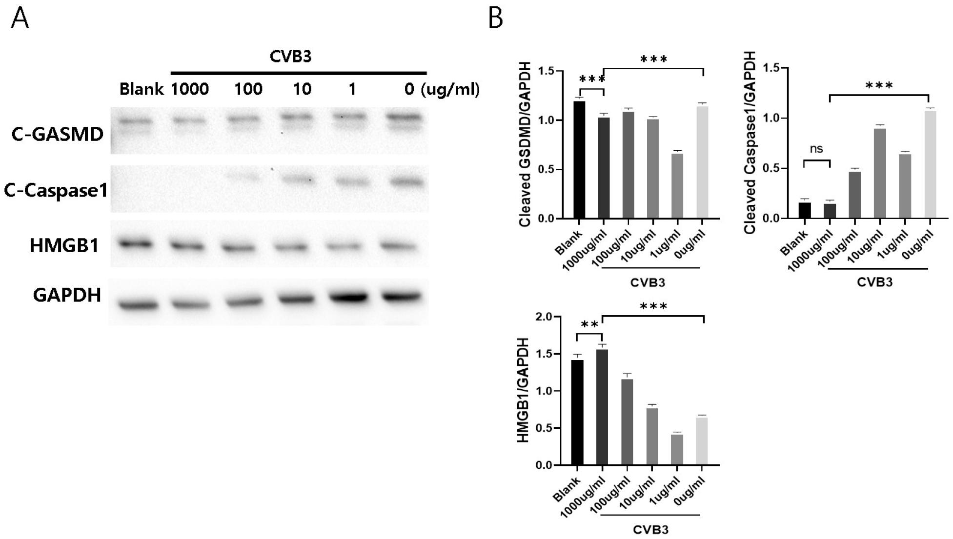
Fig. 4
H. cordata extract comprehensively suppresses the CVB3-induced pyroptotic axis by inhibiting caspase-1 activation, gasdermin D cleavage, and HMGB1 release. HeLa cells were mock-infected (Blank), infected with CVB3 alone (0 µg/mL), or infected and treated with H. cordata extract (1000-1 µg/mL). Western blot analysis was performed at 18 h post-infection. (A) Representative Western blot images showing the protein expression levels of cleaved GSDMD, cleaved caspase-1, and HMGB1. GAPDH served as the loading control. (B) Densitometric quantification of the Western blot bands relative to GAPDH. Cleaved caspase-1 and cleaved GSDMD (GSDMD-N, the pore-forming domain) were robustly induced by CVB3 infection but were significantly suppressed by H. cordata treatment in a dose-dependent manner. Conversely, intracellular HMGB1 levels were severely depleted in untreated infected cells, indicating pyroptotic release, but were dose-dependently preserved by the extract. Data are presented as the mean ± SD of three independent experiments. ns, not significant; *P < 0.05, P < 0.01, ***P < 0.001 vs. the indicated groups.
Next, we confirmed the cleavage of gasdermin D (GSDMD). GSDMD is cleaved by active caspase-1 during the terminal phase of pyroptosis to form holes in the cell membrane. The amount of cleaved GSDMD, which was significantly increased in the group infected only with the virus (0 μg/mL), significantly decreased after treatment with 1000 μg/mL of the extract. This suggests that the H. cordata extract inhibits caspase-1, thereby preventing the cleavage of GSDMD and the formation of cell membrane pores. HMGB1 is typically located safely inside the nucleus. However, when the cell membrane is destroyed due to apoptosis, this protein leaks out of the nucleus and acts as a powerful danger signal or damage- associated molecular pattern (DAMP). Our analysis revealed that CVB3 infection severely depleted intracellular HMGB1 pools compared to the mock-infected control, indicating active pyroptosis. In the 1000 µg/mL treatment group, intracellular HMGB1 was significantly preserved compared to the virus-only group, even exceeding the basal levels observed in the normal control. These findings demonstrate that the H. cordata extract effectively protects host cells and mitigates virus-induced inflammatory responses by comprehensively dismantling the CVB3-induced pyroptotic axis from caspase-1 activation and GSDMD cleavage to terminal membrane rupture and HMGB1 release. However, the reduction in viral replication may be a possible reason that weakens the induction of pyroptosis in the extract-treated group.
Immunofluorescence Visualization of CVB3 Replication and IL-18 Suppression by H. cordata Extract
To morphologically and spatially cross-validate the antiviral and pyroptosis-inhibiting effects of the H. cordata extract previously established at the protein level, immunofluorescence staining was performed (Fig. 5A and B). We evaluated the intracellular expression and localization of CVB3 VP1 (red fluorescence), a major viral capsid protein, and IL-18 (green fluorescence), a hallmark pro-inflammatory cytokine associated with pyroptosis. The experimental cohorts included a mock-infected control (Blank), a virus-only infected group (0 μg/mL), and CVB3-infected groups subsequently treated with the extract at varying concentrations (1000-100 μg/mL). In the mock-infected control, no red fluorescence corresponding to CVB3 VP1 was detected. Conversely, the virus-only infected group exhibited intense and widespread VP1 expression across the majority of the cell population. Notably, treatment with H. cordata extract markedly reduced both the intensity of intracellular red fluorescence and the overall number of infected cells in a striking dose-dependent manner (1000-100 μg/mL). These visual data compellingly corroborate our Western blot findings, reaffirming that the extract effectively impedes CVB3 replication and viral protein synthesis within host cells.
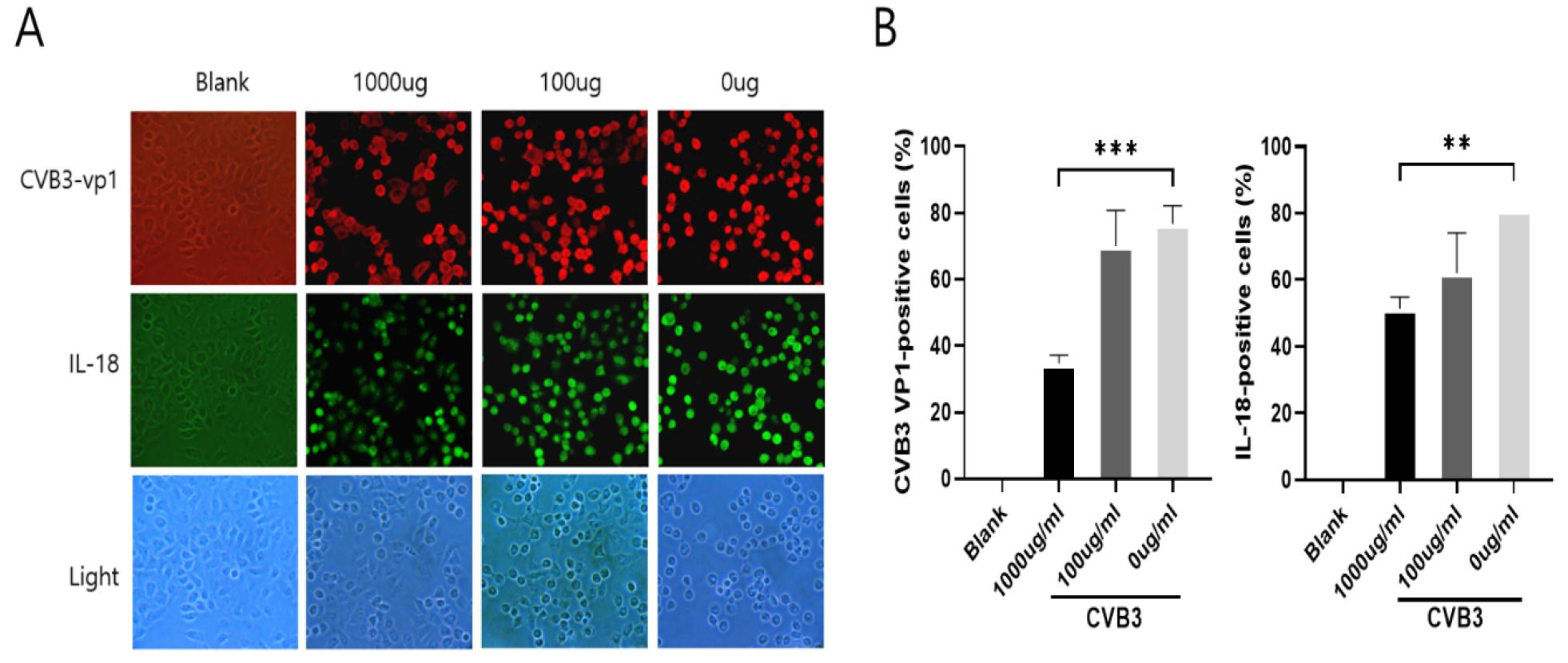
Fig. 5
H. cordata extract suppresses CVB3 replication and IL-18 expression, and prevents virus-induced morphological changes. HeLa cells were mock-infected (Blank), infected with CVB3 alone (0 µg/mL), or infected and treated with H. cordata extract (1000-100 µg/mL) for 18 h. (A) Representative immunofluorescence images showing the expression of CVB3 VP1 (red) and IL-18 (green), alongside corresponding brightfield images (bottom row) depicting cellular morphology. Untreated CVB3-infected cells exhibited intense red and green fluorescence throughout the cytoplasm, accompanied by severe cytopathic effects (cell rounding and shrinkage). Conversely, treatment with H. cordata dose-dependently reduced both VP1 and IL-18 fluorescence, with near-complete suppression at 1000 µg/mL, preserving normal cell morphology (image x200). (B) Quantitative analysis of the percentage of CVB3 VP1-positive and IL-18-positive cells. Data are presented as the mean ± SD. P < 0.001 vs. the virus-only group.
Next, we assessed the expression of IL-18, a classical inflammatory cytokine that undergoes maturation and secretion via active caspase-1 during pyroptosis. Driven by robust pyroptosis induction, the virus-only infected group displayed intense IL-18-associated green fluorescence. In stark contrast, treatment with the H. cordata extract dose-dependently attenuated this green fluorescent signal. Specifically, the 1000 μg/mL treatment group demonstrated a profound suppression of IL-18 expression compared to untreated infected cells. Furthermore, brightfield evaluation of the corresponding fields of view (bottom panels) revealed severe cellular morphological alterations in the virus-only infected group. These cells exhibited typical virus-induced cytopathic effects (CPE), hallmarked by cellular rounding, shrinkage, and the loss of cell-to-cell adhesion. Strikingly, therapeutic intervention with 1000 μg/mL of the H. cordata extract largely abrogated these morphological hallmarks of CPE, preserving a cellular architecture highly comparable to that of the mock-infected control. These results demonstrate that the H. cordata extract not only directly restricts viral replication (CVB3-VP1) but also effectively mitigates the ensuing infection-induced pyroptosis and hyperinflammatory responses (IL-18), thereby conferring both morphological and functional cytoprotection to the host cells.
DISCUSSION
Our study demonstrated a significant advancement in the search for effective therapeutic agents against Coxsackievirus B3 (CVB3) induced viral myocarditis. By demonstrating that Houttuynia cordata (H. cordata) extract effectively inhibits CVB3 replication while simultaneously suppressing the pyroptotic pathway, our findings provide novel mechanistic insights into how natural products can offer therapeutic benefits. The H. cordata extract achieves this through comprehensive inhibition of the CVB3-induced pyroptotic axis encompassing caspase-1 activation, GSDMD cleavage, and prevention of membrane rupture and HMGB1 release (15). This multi-targeted approach addresses a critical gap in current therapeutic strategies, as most existing interventions focus on single-pathway modulation rather than coordinated suppression of both viral replication and pathological immune responses. Our findings showed that the potential value of plant-derived compounds in developing safer, more efficacious treatments for CVB3-associated myocarditis and related inflammatory diseases.
Viral myocarditis represents a significant clinical challenge, particularly in children and young adults, where CVB3 remains the primary causative agent of severe myocardial inflammation. Critically, there are currently no approved targeted therapies in clinical practice that directly inhibit CVB3 replication. The pathological cascade involves primary virus-mediated cytotoxicity followed by secondary hyperimmune responses with massive infiltration of macrophages and T cells, which, if unresolved, progresses to dilated cardiomyopathy and fatal heart failure (12, 26, 27). CVB3-induced intracellular stress stimulates NLRP3 inflammasome assembly, triggering autoproteolytic caspase-1 activation, which processes pro-IL-1β and pro-IL-18 into their active forms and cleaves GSDMD. The liberated GSDMD-N oligomerizes at the plasma membrane to form 10–15 nm pores, facilitating explosive release of mature IL-1β, IL-18, and damage-associated molecular patterns (DAMPs), culminating in osmotic imbalance and pyroptotic cell death (15, 16). This pyroptotic pathway represents a highly promising strategy for attenuating excessive myocardial inflammation and preserving host cell survival in CVB3 infection. However, the reduction in viral replication may be a possible reason that weakens the induction of pyroptosis in the extract-treated group.
The development of antiviral therapeutics against CVB3 has increasingly shifted toward exploration of natural products derived from medicinal plants, reflecting both the therapeutic potential and safety advantages of plant-based compounds. Over the past two decades, numerous natural compounds have been systematically investigated for anti-CVB3 activity. Quercetin, a flavonoid abundantly found in various plant species, has demonstrated antiviral activity by inhibiting viral protease 3C and disrupting viral replication (18, 28). Resveratrol, a polyphenolic compound from grapes, has attenuated CVB3-induced myocardial injury through modulating oxidative stress and inflammatory signaling. Traditional medicinal plants have shown modern phytochemical screening, revealing multi-mechanism antiviral agents (25, 29, 30, 31). The recent study advances this research by providing detailed mechanistic evidence that Houttuynia cordata extract effectively inhibits CVB3 replication through blocking the pyroptosis signaling pathway.
In conclusion, this study provides compelling evidence that H. cordata extract is a promising natural therapeutic candidate for managing CVB3-induced viral myocarditis, with inhibition of viral replication and pyroptosis-mediated inflammation. The extract demonstrates a highly favorable safety profile at concentrations up to 1000 µg/mL. It also exhibits a potent antiviral mechanism by blocking CVB3 VP1 protein synthesis while preserving host translational machinery and abrogating infection-induced pyroptosis. By inhibiting caspase-1 activity, the H. cordata extract effectively inhibits the CVB3-induced pyroptotic axis from inflammasome assembly through terminal membrane rupture and HMGB1 release. However, this study did not prove direct mechanism for the antiviral effects of H. cordata extract. H. cordata treatment attenuates markers associated with pyroptosis rather than definitively proving direct inflammasome inhibition. In our study, direct inflammasome assays are not fully provided. Future investigations should focus on: 1) identification and isolation of active chemical constituents within H. cordata extract using advanced chromatographic and spectroscopic techniques; 2) structure-activity relationship studies to optimize compound efficacy and potency; 3) elucidation of detailed molecular mechanisms of interaction with host cell signaling pathways, viral protease targets, and inflammasome components. By pursuing these research directions, H. cordata and related natural products are anticipated to contribute substantially to the development of safe, effective, and economically accessible therapeutics for CVB3-induced viral myocarditis, fulfilling a critical unmet clinical need.