Review Article

JOURNAL OF BACTERIOLOGY AND VIROLOGY. 31 December 2024. 247-272
https://doi.org/10.4167/jbv.2024.54.4.247

ABSTRACT


MAIN

INTRODUCTION

Salmonella, a causative agent of salmonellosis in several hosts, belongs to the family Enterobacteriaceae, class Gammaproteobacteria (γ-proteobacteria), and phylum Pseudomonadota (Proteobacteria), and is a gram-negative, flagellated, facultative anaerobic, rod-shaped bacterium (1, 2). Salmonella species are primarily classified as follows: enterica and bongori (Table 1) (3). S. enterica comprises more than 2,600 serovars and is taxonomically classified into six subspecies. Moreover, DNA homology studies have revealed that S. enterica is the most clinically significant isolate (4, 5). The six phylogenetic groups of S. enterica include S. enterica subspecies enterica (I), salamae (II), arizonae (IIIa), diarizonae (IIIb), houtenae (IV), and indica (VI) (6, 7, 8, 9, 10). Among these six subspecies, the subspecies enterica is pathogenic and contains approximately 1,580 serovars, which have adverse health effects on homeotherms (11). The other subspecies are economically relevant in poikilotherms, and their pathogenicity is limited (12).

Table 1.

Salmonella species, subspecies, number of serovars, and common habitats. There are two major species of Salmonella, enterica and bongori, and over than 2,600 serovars, according to the Kauffman-White classification (12)

Species Subspecies Number of serovars Common habitats References
S. entericaenterica 1,586 Homeotherms, environments (6)
salamae 522 Poikilotherms, environments (7)
arizonae 102 Poikilotherms, environments (8)
diarizonae 338 Poikilotherms, environments (9)
houtenae 76 Poikilotherms, environments (10)
indica 13 Poikilotherms, environments (6)
S. bongori - 22 Poikilotherms, environments (3)

Salmonellosis, one of the most common foodborne diseases, impacts the health of several hosts, including animals, birds, fish, and humans, through the consumption of contaminated water and food. Based on its surface antigen composition, Salmonella subspecies I is divided into typhoidal and non-typhoidal Salmonella (13). Typhoidal Salmonella spp. (S. enterica subspecies enterica serovars Typhi and Paratyphi) have adapted to their human hosts, wherein they cause systemic infections known as typhoid and paratyphoid fever, respectively. These species are highly adapted to humans and do not cause diseases in non-human hosts (14, 15, 16, 17). Non-typhoidal Salmonella have a broad host range and most often cause gastroenteritis in humans (18). Host immunity plays a central role in shaping the course of diseases caused by non-typhoidal Salmonella(19).

Salmonella can colonize nearly all animals, including poultry, reptiles, rodents, domestic animals, birds, and humans. Animal-to-animal spread and use of Salmonella-contaminated feed maintain animal reservoirs. The most common sources of human infections include poultry, eggs, dairy products, and foods prepared on contaminated work surfaces (20). According to the Centers for Disease Control and Prevention (CDC), Salmonella is responsible for approximately 1.35 million infections, 26,500 hospitalizations, and 420 deaths annually in the United States, resulting in an estimated $400 million of direct medical costs (21). Furthermore, the Korea Disease Control and Prevention Agency (KDCA) reported an average of 2,315.78 cases of Salmonella infections over the past decade (22). Recently, the World Health Organization (WHO) updated its priority list of bacterial pathogens, identifying certain critical priority pathogens that continue to pose a major global threat. Seven years since its publication, Salmonella (S. Typhi and non-typhoidal Salmonella) has been included in the updated list as a high-priority pathogen owing to its particularly high burden in low- and middle-income countries and increasing levels of resistance (23). Despite extensive studies on Salmonella pathogenicity and infection mechanisms, salmonellosis outbreaks continue to occur worldwide, posing a threat to public health.

This review focuses on non-typhoidal Salmonella and elaborates on the causes of its infection, pathogenic mechanisms, and salmonellosis, which is the most frequent foodborne disease in humans. In addition, strategies have been described for the prevention and control of Salmonella to enhance human safety and public health.

NON-TYPHOIDAL SALMONELLA INFECTION AND PATHOGENIC MECHANISMS

Non-typhoidal Salmonella, such as S. enterica serovar Typhimurium (S. Typhimurium) is most often acquired through the consumption of contaminated food and water (17, 24, 25). However, exposure to reptiles and amphibians, which often carry bacteria, poses a risk of salmonellosis in humans. After ingestion and passage through the stomach, Salmonella attach to the mucosa of the small intestine and invade microfold (M) cells located in Peyer’s patches and enterocytes (Fig. 1) (26, 27). The bacteria remain in endocytic vacuoles, called Salmonella-containing vacuoles (SCV), wherein they replicate. Bacteria can also be transported across the cytoplasm and released into blood or lymphatic circulation (28, 29). The presence of multiple mechanisms for crossing the cellular barrier indicates the importance of this strategy in Salmonella lifecycle.
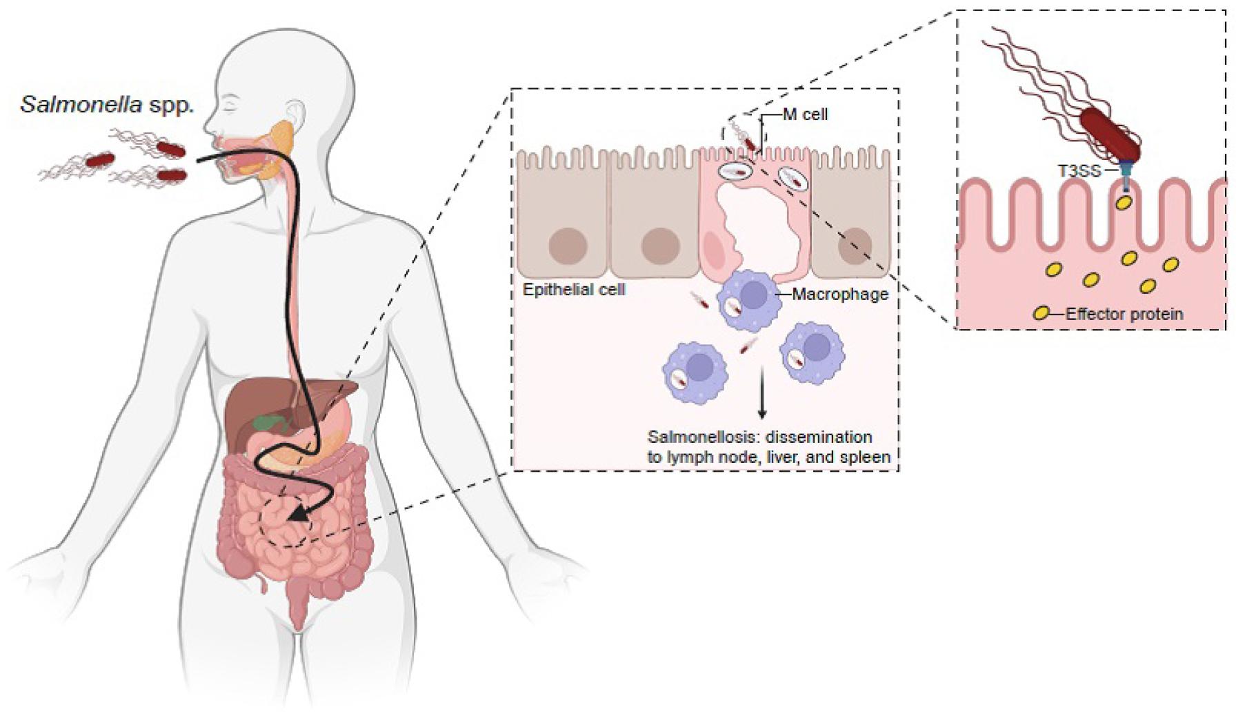
https://cdn.apub.kr/journalsite/sites/jbv/2024-054-04/N0290540401/images/JBV_2024_v54n4_247_f001.jpg
Fig. 1

Salmonella spp. mechanism of infection. Orally ingested Salmonella spp. survive at a low pH in the stomach and evade multiple defenses of the small intestine to gain epithelial access. Salmonella spp. preferentially enter M cells in the underlying Peyer’s patches. Salmonella spp. deliver effector proteins to host cells via the type III secretion system (T3SS). T3SS is encoded by Salmonella spp. within its pathogenicity island-1 (SPI-1). Effector proteins delivered by this system trigger host cell responses that result in bacterial internalization and the formation of Salmonella-containing vacuoles (SCV). Salmonella spp. induce an early local inflammatory response and salmonellosis (Created by BioRender).

Although non-typhoidal and typhoidal Salmonella share a high degree of homology, with their genomes being approximately 89% identical and having similar pathogenic mechanisms, there are a number of differences between them (30, 31). Typhoidal Salmonella harbors unique virulence factors, which are absent in non-typhoidal Salmonella(30, 31). For example, SPI-7 encodes genes for the synthesis and export of Vi capsular polysaccharide (ViCPS) (32, 33, 34), which masks lipopolysaccharide (LPS) and flagellin in S. Typhi, thereby preventing the host’s innate immune response (33, 35). Moreover, typhoidal Salmonella possesses a typhoid toxin, which is crucial for its pathogenesis and is absent in non-typhoidal Salmonella(36, 37). Some non-typhoidal Salmonella strains have typhoid toxin homologs with 1% amino acid variation, resulting in minimal systemic toxicity but preferential binding to glycan receptors on small intestinal epithelial cells (38). This corresponds to the observation that non-typhoidal Salmonella primarily localizes to the small intestine in healthy individuals, whereas typhoidal Salmonella disseminates systemically.

Pathogenicity islands (PAIs) are genetic islands related to pathogenicity, acquired by pathogenic bacteria via horizontal gene transfer. PAIs do not occur in non-pathogenic bacteria, indicating their importance in bacterial pathogenicity (39). PAIs in Salmonella spp. are called Salmonella pathogenicity islands (SPIs), and contain several genes related to Salmonella pathogenicity. To date, 17 SPIs have been identified (39). SPI-1 is a crucial factor responsible for the interactions between the host and Salmonella spp. SPI-1 is responsible for the formation of the needle-shaped type III secretion system (T3SS), which facilitates the translocation of virulence factors into host cells (Fig. 1) (40). SPI-2 also synthesizes T3SS for the translocation of effector proteins; however, the genes encoding T3SS differ from those encoding the T3SS of SPI-1 (28). SPI-3 is a 17 kb locus that contains the mgtCB operon (magnesium transport system, important for survival in nutrient starved states) and MisL, which functions as an adhesin (39, 41, 42). SPI-4 encodes genes required for the type I secretion system (T1SS) and non-fimbrial adhesin SiiE, which are involved in epithelial cell binding (43). SPI-5 is approximately 7.6 kb and encodes the effector proteins SPI-1 and -2 (44). SPI-6, also known as the Salmonella centisome 7 genomic island (SCI), contains the fimbriae-encoding gene saf and invasion protein gene pagN(45). SPI-7 is a large genetic island (134 kb) that carries genes for virulence factors, such as the Vi antigen, sopB, and type IVB pili (46). SPI-8 has been identified in S. Typhi, and encodes virulence factors with unknown functions (39). SPI-9 is involved in the adherence of S. Typhi but not in biofilm formation (47). SPI-10 is a hypervariable locus inserted into the leuX tRNA gene that encodes genes related to fimbrial adhesion, such as sef and pef(48). SPI-11 and -12 were detected in S. Choleraesuis. They exhibit the characteristics of bacteriophage genomes and tRNA genes; however, their exact functions remain unknown (49). SPI-13 is in close proximity to pheV tRNA, whereas SPI-14 is not. Both SPIs are present in S. Typhimurium and S. Enteritidis, but not in S. Typhi and S. Paratyphi (50). SPI-15, -16, and -17 are present in S. Typhi and associated with tRNA genes. Additionally, SPI-16 and -17 are also associated with LPS modulation (51).

Among the various SPI loci, SPI-1 and -2 have been extensively studied (Fig. 2) (17, 25, 40, 52). SPI-1 encompasses several operons including inv-spa, sicA-sip, prg, and org(40, 53, 54). SPI-1 also encodes T3SS, an invasion island in all Salmonella species and subspecies, with genes for invading non-phagocytic cells. Contact with intestinal epithelial cells activate T3SS, thereby delivering several effector proteins, which have the capacity to modulate various host processes (55, 56). The injected effector proteins induce cytoskeletal rearrangement, which allows the internalization of Salmonella on the epithelial cell membranes into the cellular mucosa (57). During internalization, Salmonella enters the vacuolar compartment known as SCV. The bacterium blocks the fusion of SCV with a terminal acidic lysosome, which constitutes an important intracellular defense strategy for the host cell (58). Upon sensing the phagosomal environment, SPI-2, which encodes T3SS, enables the bacterium to survive in phagocytic cells and replicate within the SCV in host cells (59, 60). Inside phagocytic cells (that is, macrophages), SCV matures, ruptures, and disseminates Salmonella into reticuloendothelial cytosol (liver and spleen) through the blood or lymphatic circulation and induces a systemic phase of infection (61). Other SPIs are primarily involved in host cell survival, replication, and production of proteins, adhesins, toxins, and fimbriae (39, 62, 63). All these effects could contribute to intestinal Salmonella colonization or intracellular growth. However, typhoidal and non-typhoidal Salmonella share T3SS, but there are differences in their pathogenesis. The role of SPI-2 in typhoidal Salmonella remains unclear, although some effectors are crucial for competitive growth in human macrophages (64, 65). Typhoidal Salmonella has a reduced functional effector repertoire, wherein approximately half of the Salmonella T3SS effectors are either pseudogenes or missing (31). The absence of the protease effector GtgE in S. Typhi, which facilitates S. Typhimurium growth in mouse macrophages by cleaving the host cell Rab32, contributes to host restriction (66). Furthermore, the SPI-1 effector SptP, essential for host cytoskeleton recovery post-infection in S. Typhimurium, is nonfunctional in S. Typhi due to variations in the SicP binding domain that impair SptP-SicP binding and hinder its translocation and activity (67, 68).
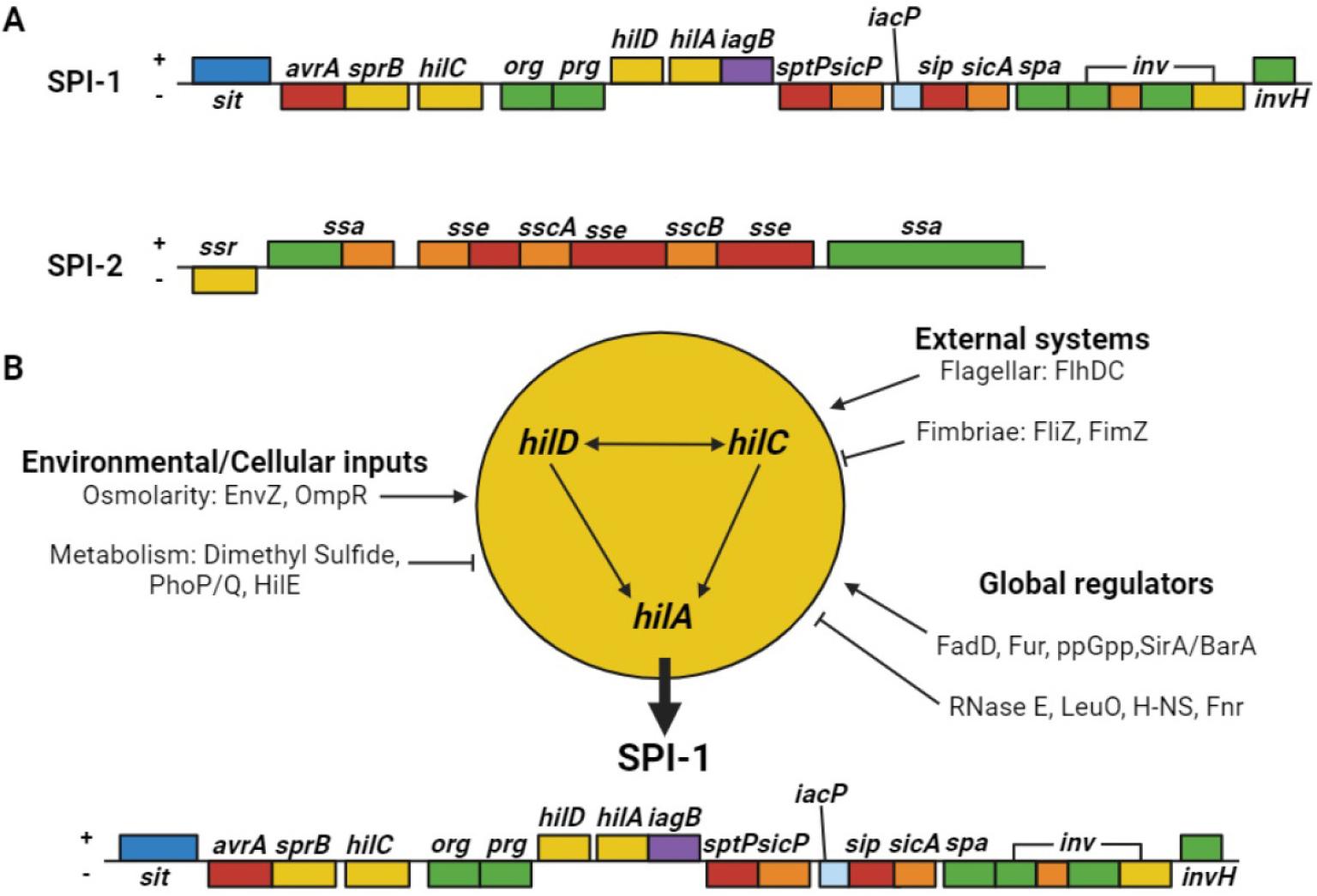
https://cdn.apub.kr/journalsite/sites/jbv/2024-054-04/N0290540401/images/JBV_2024_v54n4_247_f002.jpg
Fig. 2

Salmonella pathogenicity islands-1 and -2 (SPI-1 and -2) and associated factors. (A) Schematic illustrations of SPI-1 and -2. Each colored box represents an operon based on protein function. Blue, iron transport protein; red, T3SS secreted protein; yellow, transcriptional regulator; green, secretion apparatus; purple, cell invasion protein; orange, chaperone; light blue, likely acyl carrier protein. (B) Multiple SPI-1 gene regulators. 1) Environmental/cellular inputs: the osmolarity-related proteins EnvZ and OmpR positively regulate SPI-1, whereas metabolic inputs, such as dimethyl sulfide, PhoP/Q, and HilE, negatively regulate SPI-1. 2) External systems: FlhDC positively regulates SPI-1, whereas the fimbriae proteins FliZ and FimZ negatively regulate SPI-1 expression. 3) Global regulators: the positive regulators include FadD, Fur, ppGpp, and SirA/BarA, whereas the negative regulators include RNase E, LeuO, H-NS, and Fur.

In summary, the acquisition of broad virulence factors and functional effectors observed in non-typhoidal Salmonella appear to be consistent with its lifestyle and broad host range.

CLINICAL DISEASES CAUSED BY NON-TYPHOIDAL SALMONELLA

Globally, non-typhoidal Salmonella affects approximately 93.8 million people and causes 160,000 fatalities annually (69, 70). The primary route of Salmonella infection in humans is through the consumption of contaminated foods, including beef, pork, chicken, eggs, milk, fruits, vegetables, and water, or fecal–oral transmission (71). Gastroenteritis, septicemia, enteric fever, and asymptomatic colonization are mainly caused by Salmonella.

Gastroenteritis

Gastroenteritis is the most common form of salmonellosis worldwide (72). The onset of symptoms typically occurs 6–48 h after consuming contaminated food or water. Initially, affected individuals experienced nausea, vomiting, and non-bloody diarrhea. Fever, abdominal cramps, myalgia, and headaches are also common symptoms. Colonic involvement is observed in acute forms of the disease. Symptoms typically persist for up to 2–7 days before clearing up on their own (73).

Septicemia

All Salmonella species can cause bacteremia, although infection with typhoidal Salmonella commonly leads to a bacteremia phase. However, approximately 5% of individuals with gastrointestinal illnesses caused by non-typhoidal Salmonella develop bacteremia (18). The risk of Salmonella bacteremia is high in pediatric and geriatric patients, and immunocompromised patients (for example, those with HIV infections, sickle cell disease, and congenital immunodeficiencies) (18, 74, 75). The clinical presentation of Salmonella bacteremia is similar to that of other gram-negative bacteria; however, localized suppurative infections (for example, osteomyelitis, endocarditis, and arthritis) can occur in up to 10% of patients (76).

Enteric fever

S. Typhi causes a febrile illness known as typhoid fever. The intermediate form of this disease, referred to as paratyphoid fever, is caused by S. Paratyphi (77). Non-typhoidal Salmonella rarely cause similar symptoms (78). The bacteria responsible for enteric fever pass through the cells lining the intestine and are engulfed by macrophages (79). They replicate after translocating to the liver, spleen, and bone marrow (61). Up to 10–14 days after ingesting food or water contaminated by bacteria, patients experience a gradually increasing fever with non-specific complaints of headaches, myalgia, malaise, and anorexia. These symptoms typically persist for one week or longer and are followed by gastrointestinal symptoms. This cycle corresponds to an initial bacteremia phase, followed by gallbladder colonization, and intestine reinfection (77, 79). Enteric fever is a serious clinical disease that should be considered in febrile patients who have recently traveled to developing countries, where the disease is endemic (80).

Asymptomatic colonization

Salmonella spp. responsible for causing typhoid and paratyphoid fever are maintained through human colonization (81). Chronic colonization for more than one year after symptomatic disease develop in 3–5% of patients, with the gallbladder being the reservoir in most patients. Chronic colonization by other Salmonella spp. that cause gastroenteritis occurs in ≤ 1% of patients and does not represent an important source of human infections (82).

MULTIDRUG-RESISTANT NON-TYPHOIDAL SALMONELLA

In the 1980s, non-typhoidal Salmonella spp. were drug-sensitive. However, in the 1990s, the clinical isolates investigated at CDC exhibit increased resistance to ampicillin, chloramphenicol, and trimethoprim-sulfamethoxazole (TMP-SMZ) (18). Many countries have used various antibiotics to treat salmonellosis, leading to the emergence of antibiotic-resistant Salmonella.

Antibiotic resistance is a global concern in Salmonella spp. infections (83). In addition, it has continuously increased, exhibiting an escalating trend of 20–30% per decade. Multidrug-resistant (MDR) non-typhoidal Salmonella spp. can cause life-threatening diseases that cannot be treated with common antibiotics. Notably, MDR serotypes with high prevalence frequently tend to develop resistance against commonly prescribed antibiotics (83). MDR Salmonella spp. have also been identified in several studies. For example, MDR Salmonella was detected in 17% of broiler chickens in Egypt, with the highest resistance against neomycin (100%), nalidixic acid, cefoxitin (95%), and chloramphenicol (40.9%) (84). In 2014, the U.S. Food and Drug Administration (FDA) and a segment of the National Antimicrobial Resistance Monitoring System (NARMS) collected chicken breast samples during routine monitoring. The samples were tested using whole-genome sequencing (WGS), and MDR S. Infantis was identified, with the highest resistance observed against aminoglycosides, chloramphenicol, β-lactams, and tetracyclines. This strain includes a combination of 6–10 resistance genes (blaCTX-M-65, aac(3)-IVa, aadA1, aph(4)-Ia, aph(3′)-Ia, floR, sul1, dfrA14, tetA, and fosA) that are not common among Salmonella isolates from chickens in the United States (85). Furthermore, in the Salmonella isolates collected from 2015 to 2017, 18% of the samples were MDR Salmonella, resistant against ciprofloxacin, azithromycin, ceftriaxone, ampicillin, and TMP-SMZ (Table 2) (21, 86). Moreover, MDR Salmonella strains are also resistant to colistin, carbapenem, and tigecycline (87).

Table 2.

Drug-resistant non-typhoidal Salmonella. Some non-typhoidal Salmonella spp. become less susceptible to essential antibiotics, thereby jeopardizing the treatment of severe infections (86)

Percentage of all non-typhoidal Salmonella* Estimated number of infections per year Estimated infections per 100,000 U.S. population
Ceftriaxone resistance 3% 41,000 10
Ciprofloxacin non-susceptible 7% 89,200 30
Decreased susceptibility to azithromycin 0.5% 7,400 ≤ 5
Resistant to at least one essential antibiotic 16% 212,500 70
Resistant to three or more essential antibiotics 2% 20,800 10

Antibiotic susceptibility describes the sensitivity of bacteria to specific antibiotics. Antibiotics inhibit the growth or kill susceptible bacteria.

*Average (2015–2017)

Ciprofloxacin non-susceptible, decreased susceptibility to azithromycin, resistance to ceftriaxone, ampicillin, or trimethoprim-sulfamethoxazole.

Taken together, new therapeutic strategies, such as targeting resistance mechanisms, are required to combat MDR Salmonella development. Sequential antibiotic therapy and antibiotic rotation, both of which reduce side effects, have gained increasing attention in recent years. Chloramphenicol has typically been used to treat Salmonella infections. However, the preferred antibiotics include ampicillin, third-generation quinolones such as ciprofloxacin and levofloxacin, and third-generation cephalosporins such as ceftriaxone and macrolides (88). However, these approaches may favor cross-resistance to different antibiotics (89). Cross-resistance evaluation of different strains of Salmonella under sequential antibiotic treatment showed that effective therapeutic strategies, appropriate selection, and antibiotic therapy can help in the prevention of MDR Salmonella infections (87). Therefore, tracking the origin and development of MDR Salmonella via the implementation of nationwide monitoring systems through extensive genomic studies is crucial.

PREVENTION AND CONTROL OF NON-TYPHOIDAL SALMONELLA INFECTIONS

Several strategies are required for the prevention and control of non-typhoidal Salmonella infections in humans. Among the various strategies, the following can be considered for the prevention and control of non-typhoidal Salmonella infections.

Biosecurity measures

Biosecurity measures, including the identification of the source and transmission route (90, 91), isolation of ill animals, access control, and stringent sanitation (92, 93), prevent non-typhoidal Salmonella infections in farms. External measures include boundary fencing, vehicle regulation, and controlled animal introduction (94, 95), whereas internal measures include changing clothing and shoes, quarantining symptomatic animals, and frequent cleaning (96). High-risk visitors and uninformed farmers pose considerable risk (97, 98). Risk reduction guidelines involve limiting visitor access (99), providing clean clothing and footwear (100), removing debris, using disinfectant footbaths (101), prioritizing healthy and young animals (102), using separate or sanitized tools for food and waste, avoiding tool sharing, and restricting and sanitizing vehicles (98, 103).

Vector control and eradication

Insects, rodents, and wild birds act as vectors to facilitate the spread and persistence of non-typhoidal Salmonella infections, necessitating regular disinfection and rodent control to prevent transmission to livestock (104). Maintaining high sanitation levels is key to deterring rodents and managing insect carriers (105). Regular use of synthetic insecticides and organophosphates is recommended, although they are highly toxic and require careful application (93, 106). Natural alternatives, such as essential oils and bioinsecticides are effective, eco-friendly, and economical (107).

Isolation and quarantine

Isolation and quarantine measures effectively prevent non-typhoidal Salmonella infections and environmental contamination (108). Quarantine prevents the transmission of infection to healthy humans and protects vulnerable individuals in hospitals. Isolation units require separation from healthy areas and the proper disposal of manure (109). Quarantine duration varies based on pathogen type and exposure status (110), matching the pathogen’s incubation period for healthy exposed individuals and relying on symptoms and laboratory confirmation for infected humans (111). These measures could safeguard human health.

Hazard Analysis and Critical Control Point principles and food safety

The globally acknowledged Hazard Analysis and Critical Control Point (HACCP) system, aligned with the ISO 22000 standards and its updated protocols, enhances food safety management by preventing pathogenic contamination. However, foodborne illnesses still exist owing to inadequate cleaning, awareness, resources, and training (112). The system assesses contamination in food chains to mitigate physical, chemical, and microbiological risks, which are crucial amid evolving pathogens, antimicrobial resistance, and environmental changes. By identifying critical control points (CCPs), HACCP is essential for managing food safety and quality (113) and benefiting stakeholders across various food sectors (114).

Animal feeds

Salmonella spp., transmitted via contaminated feed ingredients (115), include serovars S. Typhimurium, S. Montevideo, S. Hadar, and S. Tennessee (116). Ensuring animal product safety can be done by reducing Salmonella contamination by heat treatment, organic acids, and chemical preservatives (117, 118, 119). Prebiotic herbs and spices, added as dried plants, extracts, or parts, enhance animal health and performance by modulating intestinal microflora, preventing Salmonella adhesion, and promoting Lactobacillus growth (120). Their antimicrobial actions disrupt microbial cell membranes, increase leakage, and diminish the proton motive force, thereby promoting appetite, digestion, physiological health, and animal productivity (121).

Epidemiological surveillance

WGS-based Salmonella surveillance identifies and manages outbreaks that are crucial for controlling human infections from contaminated food or water (122). From 1990 to 1995, S. Enteritidis and S. Typhimurium were prevalent in Europe and the Americas, mainly from poultry, while S. Typhi predominated in Africa due to poor sanitation (123). Studies linked S. Typhimurium and S. Enteritidis to poultry, and detected S. Anatum and S. Weltevreden in beef and seafood, highlighting the role of surveillance in guiding interventions and reducing the risk of salmonellosis (124). Analyzing outbreak data facilitates the interruption of disease transmission and prevention of future occurrences. Epidemiological investigations identify contamination sources and assess preventive measures (125). Implementing HACCP practices enhances food safety across production chains, thereby significantly benefiting public health (126).

Use of bacteriophages

Bacteriophages (phages) infect bacteria and are classified as lytic or lysogenic (127, 128). They are promising alternatives to antibiotics, owing to their bacteriolytic activity, specificity, self-limiting nature, and ability of genetic manipulation (129, 130, 131). Synthetic biology and genetic engineering enhance phage antibacterial efficacy, safety, and adaptability, enabling targeted pathogen elimination while preserving beneficial microbiota (132, 133). Phage biocontrol is an eco-friendly method that enhances food safety and nutritional value (134). Experimental studies that applied bacteriophage preparations (in single or cocktail form) to experimentally infected poultry and poultry products through different modes of administration and outcomes of phage treatment are outlined in Table 3(135). However, phage applications are also challenging for food processors (136). Higher concentrations are required during application, which can potentially increase costs (136, 137, 138). Bacterial resistance to frequently used or narrow-range phages can be managed by using engineered natural phages (139).

Table 3.

Overview of experimental studies examining the use of bacteriophages to reduce Salmonella spp. in poultry and related products

Experimental model Phage Inoculation dose* Phage delivery method Outcomes References
Broiler
chicken
Three Salmonella
phages
109–11 PFU Oral Within 24 h, the bacteriophage decreased cecal colonization of S. Enteritidis and S. Typhimurium by at least 4.2 and 2.9 log CFU/ml, respectively. (198)
Leghorn chicken specific-patho
gen-free (SPF)
Three-phage
cocktail
1010 PFU Oral The bacteriophage cocktail reduced Salmonella levels in the chicken cecum when administered either one day prior to or immediately following bacterial infection, and on the subsequent days post-infection (dpi). (199)
Broiler
chicken
Three-phage
cocktail
1011 PFU Oral In the group treated with bacteriophages, the number of S. Enteritidis PT4 colony-forming units per gram of cecal content decreased by 3.5-fold. (200)
Chicken
carcasses
Salmonella spp.
phage
109 PFU/ml Spraying In two experiments, S. Enteritidis was not found, while the remaining two trials demonstrated a reduction of at least 70%. (201)
SPF chicks Salmonella spp.
phage
1.18 × 10¹¹–1.03
× 102 PFU
Oral Analysis of the cecal contents revealed a modest reduction in Salmonella levels at 3 dpi, followed by a more significant decrease at 5 dpi. From 7 dpi onwards, until the study concluded at 15 dpi, all chicks tested negative for Salmonella. (202)
Broiler chicks Mixture of
bacteriophage
2.5
× 109–7.5
× 109 PFU
Oral Treatment resulted in a decrease of S. Enteritidis in cecal contents at 12 and 24 h post-administration when compared to the untreated control groups. (203)
One-day-old
chicks
Bacteriophage

ΦCJ07
105, 107
and 109
PFU
Oral All treatments effectively reduced Salmonella colonization in the intestines of both the challenged and contact chickens. Following three weeks of treatment, 70% of contact hens administered with bacteriophage at a concentration of 109 PFU/g showed no detectable intestinal Salmonella. (204)
Seven-day old
chickens
Three different
Salmonella-specific
bacteriophages
103 PFU Spray The application of competitive exclusion combined with bacteriophage resulted in a reduction of the average S. Enteritidis cecal count to 1.6 × 10² CFU/g, which was lower than 1.56 × 10⁵ CFU/g observed in the control group. (205)
Six-week-old
chickens
S. Gallinarum
(SG)-specific
bacteriophage
106 PFU Oral Contact hens administered with the bacteriophage exhibited significantly lower mortality rates than their untreated counterparts. (206)
Broiler chicks S. Enteritidis
phage
108 PFU Oral On the 14th day of the trial, the application of bacteriophage treatments substantially decreased the occurrence of S. Enteritidis in cloacal swab samples. (207)
Broiler chicks P22hc-2, cPII and
cI-7 and Felix 0
5 × 10¹¹ PFU Oral Hens treated with bacteriophages exhibited bacterial populations in their ceca that were 0.3–1.3-fold lower when compared to the untreated control group. (208)
Ten-day old
chickens
Three lytic
phages
103 PFU Spray and
Oral
The use of bacteriophages in aerosol spray form led to a 72.7% reduction in the occurrence of S. Enteritidis infection. Furthermore, measurements of S. Enteritidis levels showed that administering phages through both coarse spray and drinking water methods reduced bacterial colonization in the intestinal tract. (209)
White Leghorn
chicks
Φ st1 1012 PFU/ml Intracloacal inoculation At 6 h post-challenge, the Salmonella count decreased to 2.9 log CFU/ml, and S. Typhimurium could not be detected at or beyond the 24 h mark. (210)
Eggs PSE5 4 × 10⁷ PFU Immersion Following phage treatment, the concentration of Salmonella reduced to 2 × 10⁶ CFU/ml. (211)
Liquid egg Pu20 108 or 109
PFU/ml
Direct
inoculation
When exposed to 4 and 25°C for 24 h, the number of viable bacteria in the experimental group decreased by as much as 1.06 and 1.12 log CFU/ml, respectively. The maximum antibacterial effectiveness reached 91.30 and 92.40% at a multiplicity of infection (MOI) of 1000. (212)
Liquid whole
egg
Two phages
(OSY-STA and
OSY-SHC)
n/a Direct
inoculation
A decrease of 1.8 and more than 2.5 log CFU/ml was observed for S. Typhimurium and S. Enteritidis, respectively. (213)
Chicken
breasts and
fresh eggs

UAB_Phi 20,
UAB_Phi78, and
UAB_Phi87
109 PFU/ml and 1 ×
1010 PFU
Soaking in
suspension
and spraying
Chicken breasts exhibited decreased Salmonella levels exceeding 1 log CFU/g. A decline of 0.9 log CFU/cm2 in Salmonella was detected on the surface of fresh eggs. (214)
Raw chicken
breast
Five Salmonella
phages
3 × 10⁸ PFU Suspension
added on
surface
When incubated at 25°C, the phage-treated group exhibited the most significant decrease in S. Enteritidis and S. Typhimurium counts, with reductions of 3.06 and 2.21 log CFU/piece, respectively. (215)
Chicken breast Two-phage
cocktails
4 × 10⁹ PFU/ml Added on
surface
The concentration of S. Enteritidis in chicken breast decreased by 2.5 log CFU/sample over a 5 h period. (216)
Chicken breasts SPHG1 and
SPHG3
8.3 log PFU Spotted The bacteriophage cocktail was administered to chicken breast samples at MOI levels of 1000 or 100, resulting in a substantial decrease in the number of viable S. Typhimurium cells. (217)
Chicken
breast meat
Four Salmonella
phage
108, 109,
and 1010
PFU/ml
Directly
added
Significant reductions in viable bacterial cell counts were observed when uncooked chicken breast specimens were exposed to a cocktail of all four bacteriophages at 4°C over a 7-day period. (218)
Chicken
breast fillets
Salmonella lytic
bacteriophage
preparation
109 PFU/ml Spraying The application of chlorine and PAA, followed by a phage spray, resulted in a decrease of Salmonella by 1.6–1.7 and 2.2–2.5 log CFU/cm2, respectively. (219)
Chicken skin Eϕ151, Tϕ7
phage suspension
109 PFU Spray Following phage treatment, the reduction in Salmonella levels was observed to be 1.38 log MPN for S. Enteritidis and 1.83 log MPN for S. Typhimurium per skin area. (220)
Chicken skin vB_StyS-LmqsSP1 2.5 × 108
PFU/cm2
Direct
addition
The application of phage to chicken skin led to a decrease of approximately 2 log units in Salmonella count. This reduction was observed from the initial 3 h and persisted throughout the week-long study conducted at 4℃. (221)
Raw chicken
meat and
chicken skin
SE-P3, P16,
P37, and P47
109 PFU Direct
inoculation
When stored at 4 and 25°C, bacteriophages effectively eliminated detectable levels of Salmonella in samples that initially contained 103 CFU/g. (222)
Chicken meat Five bacteriophages 109 PFU/ml Direct
inoculation
The application of a phage cocktail led to a 1.4 log unit decrease at 48 h when compared to the control at 10℃. (223)
Chicken meat Three lytic
bacteriophages
Ic_pst11, Is_pst22,
and Is_pst24
108, 107,
and 106
PFU/ml
Direct
addition
When phage was applied at MOIs of 100, 1000, and 10,000, the number of viable S. Typhimurium cells significantly reduced after 7 h. The observed decreases were 1.17, 1.26, and 1.31 log CFU/g, respectively. (224)
Chicken meat STGO-35-1 4 × 10⁶ PFU/ml Direct
addition
The application of bacteriophage therapy resulted in a substantial decrease in S. Enteritidis, with a 2.5 log reduction observed. (225)
Chicken frankfurters Felix O1 5.25 × 10⁶ PFU Direct
addition of liquid
Two bacteriophage variants demonstrated the ability reduced S. Typhimurium levels by 1.8 and 2.1 log units, respectively. (226)
Duck meat fmb-p1 9.9 × 10⁹ PFU Direct
inoculation
A decrease of 4.52 log CFU/cm2 in S. Typhimurium levels was observed in ready-to-eat duck meat. (227)

*PFU: plaque-forming units.

Vaccination

Vaccines induce immunity without causing diseases, effectively reducing global infection rates (140, 141, 142, 143). Their efficacy varies based on formulation, with several vaccines currently in use and under development (Table 4) (93). Ty21a and Vi capsular polysaccharide vaccines effectively prevent typhoid and paratyphoid fevers (144), with Ty21a providing cross-reactive immunity to S. Typhi and S. Paratyphi (145, 146, 147) and Vi capsular polysaccharide vaccines inducing IgG production (148). Vi-conjugate vaccines are highly effective in children (149, 150, 151). However, the differences between typhoidal and non-typhoidal Salmonella strains restrict the application of typhoid vaccine technology to non-typhoidal variants (148). Vaccine development encounters challenges, including serovar genetic diversity, risks of vaccine failure, virulence reversion, tolerance to toxoid vaccines, and low immunogenicity of OMV vaccines (152, 153, 154). Continued research is essential for developing effective vaccines against evolving pathogens and diseases.

Table 4.

Available vaccines against Salmonella spp. in humans and animals

Vaccines Targets Indications Outcomes References
Vi Conjugate
(Vi-rEPA)
S. Typhi Human The effectiveness reaches up to 90% in children aged 2–5 years. There was a rapid generation of Vi-specific IgM and IgG, accompanied by a two-log decrease in bacterial shedding. (150)
Modified live
S. Dublin vaccine
(EnterVene-d)
S. Dublin Cattle Vaccinated cows exhibited enhanced cell-mediated immunity, with a 49% increase in antibody titers. The offspring of these vaccinated cows showed a more substantial increase in antibody titers, reaching 88.56%, indicating effective vertical transmission. (228)
Ty21a S. Typhi and
S. Paratyphi B
Human The T cell response exhibited cross-reactivity and multifunctionality, accompanied by an elevation in IgA production. Compared with the control, there was a 56% increase in IgA production against S. Typhi and a 38% increase against S. Paratyphi B. (144, 147)
M01ZH09, Single dose independently
attenuating deletion
(S. Typhi
(Ty2ΔaroCΔssaV) ZH9)
S. Typhi Human Swift and substantial production of IgG and IgA antibodies accompanied by bacterial elimination in feces within 7 days following infection, without any significant symptoms. (229)
GMMA, Generalized
Modules for
Membrane Antigens
S. Typhimurium,
S. Typhi, and
S. Paratyphi A
Human Enhanced activation of peripheral blood mononuclear cells led to elevated IL-6 production. This triggered a robust production of bactericidal anti-LPS O-antigen antibodies and IgG, resulting in the total elimination of the bacteria. (230, 231)
Vi Conjugate (Vi-CRM197) S. Typhi Human Exhibited 89% effectiveness in preventing typhoid fever, with protection lasting for at least four years, and substantially elevated IgG antibody levels. (151)
S. Typhi Vi
polysaccharide
tetanus toxoid
conjugate
vaccine (Tybar)
S. Typhi Human Strong anti-Vi IgG responses were observed across all age groups, demonstrating significant protection for individuals of all ages, including children under two years old. The treatment showed an effecacy of 85% without any adverse reactions. (149)
AviPro Megan
Vac 1 + A12:E13
S. Typhimurium,
S. Enteritidis, and
S. Heidelberg
Poultry S. Enteritidis was fully eliminated within 10 days following infection, with only 6% of cases remaining positive after a second inoculation. Antibodies were not vertically transmitted. (232)
Chitosan-adjuvanted
Salmonella subunit
nanoparticle vaccine
(OMPs-F-CS NPs)
S. Enteritidis Poultry Oral administration led to an increase in TLRs and Th1 and Th2 cytokine mRNA expression, along with elevated levels of OMPs-specific IgY and IgA antibodies in both saliva and intestine. The treatment resulted in a 7-fold decrease in Salmonella shedding when compared to that in the control group. (233)
Inactivated trivalent
Salmonella enterica vaccine (Nobilis®
Salenvac T; Intervet
International B.V.,
Boxmeer, The
Netherlands)
S. Typhimurium,
S. Enteritidis,
and S. Infantis
Poultry Booster dose administration in chickens resulted in a 3.9 log rise in the average antibody titer. This was accompanied by a 2.6 log decrease in cecal excretion of S. Typhimurium and S. Enteritidis, and a subsequent 1.3 log decline in S. Infantis shedding. (234)
Poulvac® ST
(Zoetis Inc.
New Jersey, USA)
S. Typhimurium,
S. Kentucky,
S. Enteritidis,
S. Heidelberg, and S. Hadar
Poultry The liver and spleen exhibited a decrease of up to 50% in five Salmonella strains: S. Kentucky, S. Enteritidis, S. Heidelberg, S. Typhimurium, and S. Hadar. This reduction demonstrated cross-protective effects among the five bacterial variants. (235)
Autologous killed
trivalent vaccine
(Tri-Vaccine)
S. Typhimurium,
S. Enteritidis, and
S. Heidelberg
Poultry Eight days after infection, 58% of the cloacal swabs collected from infected birds showed complete elimination of the bacteria. (232)

Pro- and prebiotics

Probiotics, including lactobacilli, bifidobacteria, and other lactic acid bacteria (LAB), may prevent Salmonella colonization in the gut by competing with pathogens, stimulating immune responses, and increasing gastrointestinal pH (155, 156, 157). Specifically, Lactobacillus acidophilus inhibits Salmonella spp. by secreting lactic acid, thereby lowering the intestinal pH (158). Prebiotics are fermented food ingredients that enhance the growth or activity of beneficial bacteria in the host colon and provide health benefits through their nutraceutical and nutritional value, promotion of beneficial microbiota, resistance to digestion, and intestinal fermentation (159, 160). Prebiotics support beneficial bacteria that form biofilms on the gut epithelial cells and inhibit pathogenic adhesion (161). Thus, the consumption of prebiotic-rich foods is an effective intervention against pathogenic gut bacteria (162).

Antimicrobial peptides

Antimicrobial peptides (AMPs) are a diverse group of small peptides that are crucial to the innate immune system and function through various membrane-targeting and non-membrane-targeting mechanisms (163, 164, 165). Their broad-spectrum antimicrobial properties make them promising alternatives to antibiotics with a low likelihood of developing AMR strains (166). Numerous AMPs, such as 1018-K6, A11, BmKn-2, P7, Microcin J25, HJH-3, A3-APO, IK12, Css54, Ctx(Ile21)-Ha, AvBD7, hBD-1, hBD-2, and gallinacins, show efficacy against Salmonella and other foodborne pathogens, influencing immune regulation, growth performance, and intestinal microbiota (166, 167, 168, 169, 170, 171, 172, 173, 174, 175, 176, 177, 178, 179). However, challenges, such as bacterial resistance and potential host cell toxicity, necessitate further research to enhance bioavailability and develop cost-effective production methods for their full utilization as antibiotic alternatives.

Nanoparticles

Nanoparticles (NPs) target pathogen entry sites and bypass gastrointestinal barriers, and their efficacy is determined based on size (1–500 nm) (180, 181, 182). Polymeric NPs (50–500 nm) effectively cross the intestinal mucosal barrier and are absorbed by antigen-processing cells, thus making them ideal for oral use (183). Gold nanoparticles (AuNPs) possess size- and shape-dependent photothermal properties, enhancing delivery potential due to easy functionalization (173, 184, 185, 186, 187, 188, 189, 190, 191). Silver nanoparticles (AgNPs) effectively combat gram-negative bacteria (192, 193). Chitosan nanoparticles (CNP), tested as oral Salmonella vaccine carriers, elicit immune responses in broilers and demonstrate acid stability (182, 194). Polymeric NPs resist stomach pH variations, slowly release antigens, are degradable, aid antigen-presenting cell uptake, and act as adjuvants, thereby making them an effective drug delivery system (194). Combining NPs with antibacterial agents prevents Salmonella from spreading in food and enhances human safety. Oral NP delivery with antibiotics has revolutionized typhoid fever treatment by reducing pathogen resistance, cytotoxicity, and adverse effects, while improving antibiotic localization and modulating drug-pathogen interactions (195, 196, 197).

CONCLUSIONS

Salmonella is a major foodborne pathogen and is the leading cause of gastroenteritis in humans and animals. Salmonella-contaminated food and water pose significant risks to global health. Severe complications and higher mortality rates are common in infants, the elderly, and immunocompromised individuals, with an estimated annual economic burden of $4–11 billion annually in the U.S. S. Enteritidis and S. Typhimurium are prevalent in both humans and poultry, necessitating the prevention of contamination of animals, vegetables, and fruits.

The prevention of non-typhoidal Salmonella infection is challenging, owing to the number of serovars and regional prevalence influenced by various factors. Although S. Enteritidis and S. Typhimurium are globally widespread, other serovars prevail in specific regions. Treatment differs for non-typhoidal and typhoidal Salmonella, with frequent antibiotic use for non-typhoidal infections, potentially leading to relapse and prolonged carrier states. Treating MDR strains is complicated, and no single vaccine is effective against all forms. The evolution of drug resistance has necessitated the study of serovar-specific resistance genes, with recent resistance to various antibiotics in multiple serovars.

Improved interventions by professionals, academia, and the food-processing industry are needed to address these “biological bombs.” The strategies discussed in this review can reduce Salmonella contamination. Stringent local and international policies are required to regulate the trade of live animals, plants, and animal products. Periodic epidemiological studies should trace the prevalent serovars, identify distribution vehicles, and recommend control measures to prevent or halt the trade of contaminated food or live animals in affected countries.

Each approach has unique advantages and limitations, necessitating careful consideration and further research to determine its effectiveness, safety, and scalability. As traditional treatments such as antibiotics are less effective, adopting modern and improved strategies is essential. These strategies should involve the combined use of alternative antibiotics, enhanced biosecurity measures, and prudent use of antibiotics. A comprehensive approach is the key to controlling the spread of Salmonella. Additionally, a One Health approach and ongoing collaboration among researchers, healthcare professionals, veterinarians, and policymakers in antibiotic stewardship are vital for protecting public health and food safety from prevalent foodborne pathogens such as Salmonella.

AUTHOR CONTRIBUTION

M Lee: Conceptualization; Data curation; Formal analysis; Funding acquisition; Investigation; Project administration; Resources; Supervision; Validation; Visualization; Roles/Writing - original draft; and Writing - review & editing.

CONFLICT OF INTEREST

The author declares no conflicts of interest.

Acknowledgements

This work was supported by the National Research Foundation of Korea (NRF) grant funded by the Korea government (Ministry of Science and ICT; MSIT) (Grant No. RS-2023-00210754).

References

1

Adeolu M, Alnajar S, Naushad S, S Gupta R. Genome-based phylogeny and taxonomy of the 'Enterobacteriales' : proposal for Enterobacterales ord. nov. divided into the families Enterobacteriaceae, Erwiniaceae fam. nov., Pectobacteriaceae fam. nov., Yersiniaceae fam. nov., Hafniaceae fam. nov., Morganellaceae fam. nov., and Budviciaceae fam. nov. Int J Syst Evol Microbiol. 2016;66(12):5575-5599.

10.1099/ijsem.0.00148527620848
2

Ryan MP, O'Dwyer J, Adley CC. Evaluation of the Complex Nomenclature of the Clinically and Veterinary Significant Pathogen Salmonella. Biomed Res Int. 2017;2017:3782182.

10.1155/2017/378218228540296PMC5429938
3

Fookes M, Schroeder GN, Langridge GC, Blondel CJ, Mammina C, Connor TR, et al. Salmonella bongori provides insights into the evolution of the Salmonellae. PLoS Pathog. 2011;7(8):e1002191.

10.1371/journal.ppat.100219121876672PMC3158058
4

Brenner FW, Villar RG, Angulo FJ, Tauxe R, Swaminathan B. Salmonella nomenclature. J Clin Microbiol. 2000;38(7):2465-2467.

10.1128/JCM.38.7.2465-2467.200010878026PMC86943
5

LeLievre V, Besnard A, Schlusselhuber M, Desmasures N, Dalmasso M. Phages for biocontrol in foods: What opportunities for Salmonella sp. control along the dairy food chain? Food Microbiol. 2019;78:89-98.

10.1016/j.fm.2018.10.00930497612
6

Agasan A, Kornblum J, Williams G, Pratt CC, Fleckenstein P, Wong M, et al. Profile of Salmonella enterica subsp. enterica (subspecies I) serotype 4,5,12:i:- strains causing food-borne infections in New York City. J Clin Microbiol. 2002;40(6):1924-1929.

10.1128/JCM.40.6.1924-1929.200212037044PMC130705
7

Hounmanou YMG, Baniga Z, Garcia V, Dalsgaard A. Salmonella Salamae and S. Waycross isolated from Nile perch in Lake Victoria show limited human pathogenic potential. Sci Rep. 2022;12(1):4229.

10.1038/s41598-022-08200-535273308PMC8913728
8

Mahajan RK, Khan SA, Chandel DS, Kumar N, Hans C, Chaudhry R. Fatal case of Salmonella enterica subsp. arizonae gastroenteritis in an infant with microcephaly. J Clin Microbiol. 2003;41(12):5830-5832.

10.1128/JCM.41.12.5830-5832.2003PMC309002
9

Giner-Lamia J, Vinuesa P, Betancor L, Silva C, Bisio J, Soleto L, et al. Genome analysis of Salmonella enterica subsp. diarizonae isolates from invasive human infections reveals enrichment of virulence-related functions in lineage ST1256. BMC Genomics. 2019;20(1):99.

10.1186/s12864-018-5352-z30704413PMC6357384
10

Andruzzi MN, Krath ML, Lawhon SD, Boudreau B. Salmonella enterica subspecies houtenae as an opportunistic pathogen in a case of meningoencephalomyelitis and bacteriuria in a dog. BMC Vet Res. 2020;16(1):437.

10.1186/s12917-020-02652-533176763PMC7659121
11

Ferrari RG, Rosario DKA, Cunha-Neto A, Mano SB, Figueiredo EES, Conte-Junior CA. Worldwide Epidemiology of Salmonella Serovars in Animal-Based Foods: a Meta-analysis. Appl Environ Microbiol. 2019;85(14):e00591-19.

10.1128/AEM.00591-1931053586PMC6606869
12

Issenhuth-Jeanjean S, Roggentin P, Mikoleit M, Guibourdenche M, de Pinna E, Nair S, et al. Supplement 2008-2010 (no. 48) to the White-Kauffmann-Le Minor scheme. Res Microbiol. 2014;165(7):526-530.

10.1016/j.resmic.2014.07.00425049166
13

Smith SI, Seriki A, Ajayi A. Typhoidal and non-typhoidal Salmonella infections in Africa. Eur J Clin Microbiol Infect Dis. 2016;35(12):1913-1922.

10.1007/s10096-016-2760-327562406
14

House D, Bishop A, Parry C, Dougan G, Wain J. Typhoid fever: pathogenesis and disease. Curr Opin Infect Dis. 2001;14(5):573-578.

10.1097/00001432-200110000-0001111964878
15

Parry CM, Hien TT, Dougan G, White NJ, Farrar JJ. Typhoid fever. N Engl J Med. 2002;347(22):1770-1782.

10.1056/NEJMra02020112456854
16

Dougan G, Baker S. Salmonella enterica serovar Typhi and the pathogenesis of typhoid fever. Annu Rev Microbiol. 2014;68:317-336.

10.1146/annurev-micro-091313-10373925208300
17

Galan JE. Salmonella Typhimurium and inflammation: a pathogen-centric affair. Nat Rev Microbiol. 2021;19(11):716-725.

10.1038/s41579-021-00561-434012042PMC9350856
18

Hohmann EL. Nontyphoidal salmonellosis. Clin Infect Dis. 2001;32(2):263-269.

10.1086/31845711170916
19

Rivera-Chavez F, Baumler AJ. The Pyromaniac Inside You: Salmonella Metabolism in the Host Gut. Annu Rev Microbiol. 2015;69:31-48.

10.1146/annurev-micro-091014-10410826002180
20

Kim JH, Song SM, Kim JH, Lim SM, Park SJ, Nam HJ, et al. Trends in Acute Gastroenteritis through the Pathogen Surveillance System in Incheon Metropolitan City, 2018-2021. J Bacteriol Virol. 2022;52(2):54-63.

10.4167/jbv.2022.52.2.054
1

U. S. Centers for Disease Control and Prevention. Salmonella Homepage. Available at https://www.cdc.gov/salmonella/index.html [accessed on 3 October 2024].

2

Korea Disease Control and Prevention Agency. Desease Portal/Desease Statics/Salmonella Infection. Available at https://dportal.kdca.go.kr/pot/ii/sttyInftnsds/sttyInftnsds.do?icdCd=ND0601 [accessed on 19 August 2024].

3

Jesudason T. WHO publishes updated list of bacterial priority pathogens. Lancet Microbe. 2024;5(9):100940.

10.1016/j.lanmic.2024.07.00339079540
4

Ohl ME, Miller SI. Salmonella: a model for bacterial pathogenesis. Annu Rev Med. 2001;52:259-274.

10.1146/annurev.med.52.1.25911160778
5

Haraga A, Ohlson MB, Miller SI. Salmonellae interplay with host cells. Nat Rev Microbiol. 2008;6(1):53-66.

10.1038/nrmicro178818026123
6

Kohbata S, Yokoyama H, Yabuuchi E. Cytopathogenic effect of Salmonella typhi GIFU 10007 on M cells of murine ileal Peyer's patches in ligated ileal loops: an ultrastructural study. Microbiol Immunol. 1986;30(12):1225-1237.

10.1111/j.1348-0421.1986.tb03055.x3553868
7

Jones BD, Ghori N, Falkow S. Salmonella typhimurium initiates murine infection by penetrating and destroying the specialized epithelial M cells of the Peyer's patches. J Exp Med. 1994;180(1):15-23.

10.1084/jem.180.1.158006579PMC2191576
8

Jennings E, Thurston TLM, Holden DW. Salmonella SPI-2 Type III Secretion System Effectors: Molecular Mechanisms And Physiological Consequences. Cell Host Microbe. 2017;22(2):217-231.

10.1016/j.chom.2017.07.00928799907
9

Wotzka SY, Nguyen BD, Hardt WD. Salmonella Typhimurium Diarrhea Reveals Basic Principles of Enteropathogen Infection and Disease-Promoted DNA Exchange. Cell Host Microbe. 2017;21(4):443-454.

10.1016/j.chom.2017.03.00928407482
30

McClelland M, Sanderson KE, Spieth J, Clifton SW, Latreille P, Courtney L, et al. Complete genome sequence of Salmonella enterica serovar Typhimurium LT2. Nature. 2001;413(6858):852-856.

10.1038/3510161411677609
31

Parkhill J, Dougan G, James KD, Thomson NR, Pickard D, Wain J, et al. Complete genome sequence of a multiple drug resistant Salmonella enterica serovar Typhi CT18. Nature. 2001;413(6858):848-852.

10.1038/3510160711677608
32

Sabbagh SC, Forest CG, Lepage C, Leclerc JM, Daigle F. So similar, yet so different: uncovering distinctive features in the genomes of Salmonella enterica serovars Typhimurium and Typhi. FEMS Microbiol Lett. 2010;305(1):1-13.

10.1111/j.1574-6968.2010.01904.x20146749
33

Raffatellu M, Chessa D, Wilson RP, Dusold R, Rubino S, Baumler AJ. The Vi capsular antigen of Salmonella enterica serotype Typhi reduces Toll-like receptor-dependent interleukin-8 expression in the intestinal mucosa. Infect Immun. 2005;73(6):3367-3374.

10.1128/IAI.73.6.3367-3374.200515908363PMC1111811
34

Yang YA, Chong A, Song J. Why Is Eradicating Typhoid Fever So Challenging: Implications for Vaccine and Therapeutic Design. Vaccines (Basel). 2018;6(3):45.

10.3390/vaccines603004530042307PMC6160957
35

Wangdi T, Lee CY, Spees AM, Yu C, Kingsbury DD, Winter SE, et al. The Vi capsular polysaccharide enables Salmonella enterica serovar typhi to evade microbe-guided neutrophil chemotaxis. PLoS Pathog. 2014;10(8):e1004306.

10.1371/journal.ppat.100430625101794PMC4125291
36

Chong A, Lee S, Yang YA, Song J. The Role of Typhoid Toxin in Salmonella Typhi Virulence. Yale J Biol Med. 2017;90(2):283-290.

37

Galan JE. Typhoid toxin provides a window into typhoid fever and the biology of Salmonella Typhi. Proc Natl Acad Sci U S A. 2016;113(23):6338-6344.

10.1073/pnas.160633511327222578PMC4988619
38

Miller RA, Wiedmann M. The Cytolethal Distending Toxin Produced by Nontyphoidal Salmonella Serotypes Javiana, Montevideo, Oranienburg, and Mississippi Induces DNA Damage in a Manner Similar to That of Serotype Typhi. mBio. 2016;7(6):e02109-16.

10.1128/mBio.02109-1627999166PMC5181781
39

Kombade S, Kaur N. Pathogenicity Island in Salmonella. In: Lamas A, editor. Salmonella spp - A Global Challenge. IntechOpen; 2021.

10.5772/intechopen.96443
40

Lou L, Zhang P, Piao R, Wang Y. Salmonella Pathogenicity Island 1 (SPI-1) and Its Complex Regulatory Network. Front Cell Infect Microbiol. 2019;9:270.

10.3389/fcimb.2019.0027031428589PMC6689963
41

Retamal P, Castillo-Ruiz M, Mora GC. Characterization of MgtC, a virulence factor of Salmonella enterica Serovar Typhi. PLoS One. 2009;4(5):e5551.

10.1371/journal.pone.000555119436747PMC2677668
42

Wang S, Yang D, Wu X, Wang Y, Wang D, Tian M, et al. Autotransporter MisL of Salmonella enterica serotype Typhimurium facilitates bacterial aggregation and biofilm formation. FEMS Microbiol Lett. 2018;365(17).

10.1093/femsle/fny142
43

Gerlach RG, Jackel D, Stecher B, Wagner C, Lupas A, Hardt WD, et al. Salmonella Pathogenicity Island 4 encodes a giant non-fimbrial adhesin and the cognate type 1 secretion system. Cell Microbiol. 2007;9(7):1834-1850.

10.1111/j.1462-5822.2007.00919.x17388786
44

Knodler LA, Celli J, Hardt WD, Vallance BA, Yip C, Finlay BB. Salmonella effectors within a single pathogenicity island are differentially expressed and translocated by separate type III secretion systems. Mol Microbiol. 2002;43(5):1089-1103.

10.1046/j.1365-2958.2002.02820.x11918798
45

Folkesson A, Lofdahl S, Normark S. The Salmonella enterica subspecies I specific centisome 7 genomic island encodes novel protein families present in bacteria living in close contact with eukaryotic cells. Res Microbiol. 2002;153(8):537-545.

10.1016/S0923-2508(02)01348-712437215
46

Seth-Smith HM. SPI-7: Salmonella's Vi-encoding Pathogenicity Island. J Infect Dev Ctries. 2008;2(4):267-271.

10.3855/jidc.22019741287
47

Velasquez JC, Hidalgo AA, Villagra N, Santiviago CA, Mora GC, Fuentes JA. SPI-9 of Salmonella enterica serovar Typhi is constituted by an operon positively regulated by RpoS and contributes to adherence to epithelial cells in culture. Microbiology (Reading). 2016;162(8):1367-1378.

10.1099/mic.0.00031927260307
48

Bishop AL, Baker S, Jenks S, Fookes M, Gaora PO, Pickard D, et al. Analysis of the hypervariable region of the Salmonella enterica genome associated with tRNAleuX. J Bacteriol. 2005;187(7):2469-2482.

10.1128/JB.187.7.2469-2482.200515774890PMC1065210
49

Chiu CH, Tang P, Chu C, Hu S, Bao Q, Yu J, et al. The genome sequence of Salmonella enterica serovar Choleraesuis, a highly invasive and resistant zoonotic pathogen. Nucleic Acids Res. 2005;33(5):1690-1698.

10.1093/nar/gki29715781495PMC1069006
50

Shah DH, Lee MJ, Park JH, Lee JH, Eo SK, Kwon JT, et al. Identification of Salmonella gallinarum virulence genes in a chicken infection model using PCR-based signature-tagged mutagenesis. Microbiology (Reading). 2005;151(Pt 12):3957-3968.

10.1099/mic.0.28126-016339940
51

Vernikos GS, Parkhill J. Interpolated variable order motifs for identification of horizontally acquired DNA: revisiting the Salmonella pathogenicity islands. Bioinformatics. 2006;22(18):2196-2203.

10.1093/bioinformatics/btl36916837528
52

Lee M, Ryu M, Joo M, Seo YJ, Lee J, Kim HM, et al. Endoribonuclease-mediated control of hns mRNA stability constitutes a key regulatory pathway for Salmonella Typhimurium pathogenicity island 1 expression. PLoS Pathog. 2021;17(2):e1009263.

10.1371/journal.ppat.100926333524062PMC7877770
53

Song M, Sukovich DJ, Ciccarelli L, Mayr J, Fernandez-Rodriguez J, Mirsky EA, et al. Control of type III protein secretion using a minimal genetic system. Nat Commun. 2017;8:14737.

10.1038/ncomms1473728485369PMC5436071
54

Ellermeier JR, Slauch JM. Adaptation to the host environment: regulation of the SPI1 type III secretion system in Salmonella enterica serovar Typhimurium. Curr Opin Microbiol. 2007;10(1):24-29.

10.1016/j.mib.2006.12.00217208038
55

LaRock DL, Chaudhary A, Miller SI. Salmonellae interactions with host processes. Nat Rev Microbiol. 2015;13(4):191-205.

10.1038/nrmicro342025749450PMC5074537
56

Figueira R, Holden DW. Functions of the Salmonella pathogenicity island 2 (SPI-2) type III secretion system effectors. Microbiology (Reading). 2012;158(Pt 5):1147-1161.

10.1099/mic.0.058115-022422755
57

Zhou D, Mooseker MS, Galan JE. Role of the S. typhimurium actin-binding protein SipA in bacterial internalization. Science. 1999;283(5410):2092-2095.

10.1126/science.283.5410.209210092234
58

Eswarappa SM, Negi VD, Chakraborty S, Chandrasekhar Sagar BK, Chakravortty D. Division of the Salmonella-containing vacuole and depletion of acidic lysosomes in Salmonella-infected host cells are novel strategies of Salmonella enterica to avoid lysosomes. Infect Immun. 2010;78(1):68-79.

10.1128/IAI.00668-0919858305PMC2798212
59

Cirillo DM, Valdivia RH, Monack DM, Falkow S. Macrophage-dependent induction of the Salmonella pathogenicity island 2 type III secretion system and its role in intracellular survival. Mol Microbiol. 1998;30(1):175-188.

10.1046/j.1365-2958.1998.01048.x9786194
60

Lee AK, Detweiler CS, Falkow S. OmpR regulates the two-component system SsrA-SsrB in Salmonella pathogenicity island 2. J Bacteriol. 2000;182(3):771-781.

10.1128/JB.182.3.771-781.200010633113PMC94342
61

Waterman SR, Holden DW. Functions and effectors of the Salmonella pathogenicity island 2 type III secretion system. Cell Microbiol. 2003;5(8):501-511.

10.1046/j.1462-5822.2003.00294.x12864810
62

Klingl S, Kordes S, Schmid B, Gerlach RG, Hensel M, Muller YA. Recombinant protein production and purification of SiiD, SiiE and SiiF - Components of the SPI4-encoded type I secretion system from Salmonella Typhimurium. Protein Expr Purif. 2020;172:105632.

10.1016/j.pep.2020.10563232251835
63

Singh Y, Saxena A, Kumar R, Saxena MK. Virulence System of Salmonella with Special Reference to Salmonella enterica. In: Mascellino MT, editor. Salmonella - A Re-emerging Pathogen. IntechOpen; 2018.

10.5772/intechopen.77210
64

Forest CG, Ferraro E, Sabbagh SC, Daigle F. Intracellular survival of Salmonella enterica serovar Typhi in human macrophages is independent of Salmonella pathogenicity island (SPI)-2. Microbiology (Reading). 2010;156(Pt 12):3689-3698.

10.1099/mic.0.041624-020817644
65

Sabbagh SC, Lepage C, McClelland M, Daigle F. Selection of Salmonella enterica serovar Typhi genes involved during interaction with human macrophages by screening of a transposon mutant library. PLoS One. 2012;7(5):e36643.

10.1371/journal.pone.003664322574205PMC3344905
66

Spano S, Galan JE. A Rab32-dependent pathway contributes to Salmonella typhi host restriction. Science. 2012;338(6109):960-963.

10.1126/science.122922423162001PMC3693731
67

Fu Y, Galan JE. A Salmonella protein antagonizes Rac-1 and Cdc42 to mediate host-cell recovery after bacterial invasion. Nature. 1999;401(6750):293-297.

10.1038/4582910499590
68

Johnson R, Byrne A, Berger CN, Klemm E, Crepin VF, Dougan G, et al. The Type III Secretion System Effector SptP of Salmonella enterica Serovar Typhi. J Bacteriol. 2017;199(4): e00647-16.

10.1128/JB.00647-16
69

Balasubramanian R, Im J, Lee JS, Jeon HJ, Mogeni OD, Kim JH, et al. The global burden and epidemiology of invasive non-typhoidal Salmonella infections. Hum Vaccin Immunother. 2019;15(6):1421-1426.

10.1080/21645515.2018.150471730081708PMC6663144
70

Allen JC, Toapanta FR, Baliban SM, Sztein MB, Tennant SM. Reduced immunogenicity of a live Salmonella enterica serovar Typhimurium vaccine in aged mice. Front Immunol. 2023;14:1190339.

10.3389/fimmu.2023.119033937207226PMC10188964
71

Pui CF, Wong WC, Chai LC, Nillian E, Ghazali FM, Cheah YK, et al. Simultaneous detection of Salmonella spp., Salmonella Typhi and Salmonella Typhimurium in sliced fruits using multiplex PCR. Food Control. 2011;22(2):337-342.

10.1016/j.foodcont.2010.05.021
72

Eng SK, Pusparajah P, Ab Mutalib NS, Ser HL, Chan KG, Lee LH. Salmonella: A review on pathogenesis, epidemiology and antibiotic resistance. Front Life Sci. 2015;8(3):284-293.

10.1080/21553769.2015.1051243
73

Getto L, Zeserson E, Breyer M. Vomiting, diarrhea, constipation, and gastroenteritis. Emerg Med Clin North Am. 2011;29(2):211-237.

10.1016/j.emc.2011.01.00521515177PMC7132768
74

Zaidi E, Bachur R, Harper M. Non-typhi Salmonella bacteremia in children. Pediatr Infect Dis J. 1999;18(12):1073-1077.

10.1097/00006454-199912000-0000910608627
75

Galofre J, Moreno A, Mensa J, Miro JM, Gatell JM, Almela M, et al. Analysis of factors influencing the outcome and development of septic metastasis or relapse in Salmonella bacteremia. Clin Infect Dis. 1994;18(6):873-878.

10.1093/clinids/18.6.8738086546
76

Jiang B, Xu H, Zhou Z. Septic arthritis with osteomyelitis due to Salmonella enterica serotype Dublin: A case series. Front Surg. 2023;9:1069141.

10.3389/fsurg.2022.106914136684367PMC9852604
77

Meiring JE, Khanam F, Basnyat B, Charles RC, Crump JA, Debellut F, et al. Typhoid fever. Nat Rev Dis Primers. 2023;9(1):71.

10.1038/s41572-023-00480-z38097589
78

Wang BX, Butler DS, Hamblin M, Monack DM. One species, different diseases: the unique molecular mechanisms that underlie the pathogenesis of typhoidal Salmonella infections. Curr Opin Microbiol. 2023;72:102262.

10.1016/j.mib.2022.10226236640585PMC10023398
79

Neupane DP, Dulal HP, Song J. Enteric Fever Diagnosis: Current Challenges and Future Directions. Pathogens. 2021;10(4):410.

10.3390/pathogens1004041033915749PMC8065732
80

Basnyat B, Maskey AP, Zimmerman MD, Murdoch DR. Enteric (typhoid) fever in travelers. Clin Infect Dis. 2005; 41(10):1467-1472.

10.1086/49713616231259
81

Alvarez-Ordonez A, Begley M, Prieto M, Messens W, Lopez M, Bernardo A, et al. Salmonella spp. survival strategies within the host gastrointestinal tract. Microbiology (Reading). 2011;157(Pt 12):3268-3281.

10.1099/mic.0.050351-022016569
82

Gonzalez-Escobedo G, Marshall JM, Gunn JS. Chronic and acute infection of the gall bladder by Salmonella Typhi: understanding the carrier state. Nat Rev Microbiol. 2011;9(1):9-14.

10.1038/nrmicro249021113180PMC3255095
83

Liljebjelke KA, Hofacre CL, White DG, Ayers S, Lee MD, Maurer JJ. Diversity of Antimicrobial Resistance Phenotypes in Salmonella Isolated from Commercial Poultry Farms. Front Vet Sci. 2017;4:96.

10.3389/fvets.2017.0009628691011PMC5482141
84

Elsayed MM, El-Basrey YFH, El-Baz AH, Dowidar HA, Shami A, Al-Saeed FA, et al. Ecological prevalence, genetic diversity, and multidrug resistance of Salmonella enteritidis recovered from broiler and layer chicken farms. Poult Sci. 2024;103(2):103320.

10.1016/j.psj.2023.10332038215504PMC10825688
85

Srednik ME, Morningstar-Shaw BR, Hicks JA, Tong C, Mackie TA, Schlater LK. Whole-genome sequencing and phylogenetic analysis capture the emergence of a multi-drug resistant Salmonella enterica serovar Infantis clone from diagnostic animal samples in the United States. Front Microbiol. 2023;14:1166908.

10.3389/fmicb.2023.116690837333652PMC10272548
86

U. S. Centers for Disease Control and Prevention. Antibiotic resistance threats in the United States, 2019. Available at https://www.cdc.gov/drugresistance/Biggest-Threats.html [accessed on 16 July 2024].

87

Dawan J, Ahn J. Assessment of cross-resistance potential to serial antibiotic treatments in antibiotic-resistant Salmonella Typhimurium. Microb Pathog. 2020;148:104478.

10.1016/j.micpath.2020.10447832916245
88

Bhandari M, Poelstra JW, Kauffman M, Varghese B, Helmy YA, Scaria J, et al. Genomic Diversity, Antimicrobial Resistance, Plasmidome, and Virulence Profiles of Salmonella Isolated from Small Specialty Crop Farms Revealed by Whole-Genome Sequencing. Antibiotics (Basel). 2023;12(11):1637.

10.3390/antibiotics1211163737998839PMC10668983
89

Vestergaard M, Paulander W, Marvig RL, Clasen J, Jochumsen N, Molin S, et al. Antibiotic combination therapy can select for broad-spectrum multidrug resistance in Pseudomonas aeruginosa. Int J Antimicrob Agents. 2016;47(1):48-55.

10.1016/j.ijantimicag.2015.09.01426597931
90

Good practices for biosecurity in the pig sector: Issues and options in developing and transition countries. Available at https://www.fao.org/4/i1435e/i1435e00.htm [accessed on 30 December 2010].

91

Galipo E, Zoche-Golob V, Sassu EL, Prigge C, Sjolund M, Tobias T, et al. Prioritization of pig farm biosecurity for control of Salmonella and hepatitis E virus infections: results of a European expert opinion elicitation. Porcine Health Manag. 2023;9(1):8.

10.1186/s40813-023-00306-036872376PMC9987137
92

Sharma B. Poultry Production, Management and Bio-Security Measures. J Agric Environ. 2010;11:120-125.

10.3126/aej.v11i0.3659
93

Lamichhane B, Mawad AMM, Saleh M, Kelley WG, Harrington PJ 2nd, Lovestad CW, et al. Salmonellosis: An Overview of Epidemiology, Pathogenesis, and Innovative Approaches to Mitigate the Antimicrobial Resistant Infections. Antibiotics (Basel). 2024;13(1):76.

10.3390/antibiotics1301007638247636PMC10812683
94

Jensen AN, Dalsgaard A, Stockmarr A, Nielsen EM, Baggesen DL. Survival and transmission of Salmonella enterica serovar Typhimurium in an outdoor organic pig farming environment. Appl Environ Microbiol. 2006;72(3):1833-1842.

10.1128/AEM.72.3.1833-1842.200616517629PMC1393233
95

Mannion C, Leonard FC, Lynch PB, Egan J. Efficacy of cleaning and disinfection on pig farms in Ireland. Vet Rec. 2007;161(11):371-375.

10.1136/vr.161.11.37117873266
96

Trampel DW, Holder TG, Gast RK. Integrated farm management to prevent Salmonella Enteritidis contamination of eggs. J Appl Poult Res. 2014;23(2):353-365.

10.3382/japr.2014-00944
97

Mee JF, Geraghty T, O'Neill R, More SJ. Bioexclusion of diseases from dairy and beef farms: risks of introducing infectious agents and risk reduction strategies. Vet J. 2012;194(2):143-150.

10.1016/j.tvjl.2012.07.00123103219PMC7110757
98

Renault V, Damiaans B, Sarrazin S, Humblet MF, Dewulf J, Saegerman C. Biosecurity practices in Belgian cattle farming: Level of implementation, constraints and weaknesses. Transbound Emerg Dis. 2018;65(5):1246-1261.

10.1111/tbed.1286529566303
99

Tilli G, Laconi A, Galuppo F, Mughini-Gras L, Piccirillo A. Assessing Biosecurity Compliance in Poultry Farms: A Survey in a Densely Populated Poultry Area in North East Italy. Animals (Basel). 2022;12(11):1409.

10.3390/ani1211140935681871PMC9179503
100

Busani L, Dalla Pozza M, Bonfanti L, Toson M, Ferre N, Marangon S. Intervention strategies for low-pathogenic avian influenza control in Italy. Avian Dis. 2007;51(1 Suppl):470-473.

10.1637/7553-033106R.117494610
101

Gelaude P, Schlepers M, Verlinden M, Laanen M, Dewulf J. Biocheck.UGent: a quantitative tool to measure biosecurity at broiler farms and the relationship with technical performances and antimicrobial use. Poult Sci. 2014;93(11):2740-2751.

10.3382/ps.2014-0400225193257
102

Van Limbergen T, Dewulf J, Klinkenberg M, Ducatelle R, Gelaude P, Mendez J, et al. Scoring biosecurity in European conventional broiler production. Poult Sci. 2018;97(1):74-83.

10.3382/ps/pex29629077940
103

Maunsell F, Donovan GA. Biosecurity and risk management for dairy replacements. Vet Clin North Am Food Anim Pract. 2008;24(1):155-190.

10.1016/j.cvfa.2007.10.00718299037PMC7134781
104

Verwoerd DW. Definition of a vector and a vector-borne disease. Rev Sci Tech. 2015;34(1):29-39.

10.20506/rst.34.1.234326470447
105

Axtell RC. Fly Management in Poultry Production: Cultural, Biological, and Chemical. Poult Sci. 1986;65(4):657-667.

10.3382/ps.0650657
106

Balaraman V, Drolet BS, Mitzel DN, Wilson WC, Owens J, Gaudreault NN, et al. Mechanical transmission of SARS-CoV-2 by house flies. Parasit Vectors. 2021;14(1):214.

10.1186/s13071-021-04703-833879234PMC8056201
107

Azizi-Lalabadi M, Rahimzadeh-Sani Z, Feng J, Hosseini H, Jafari SM. The impact of essential oils on the qualitative properties, release profile, and stimuli-responsiveness of active food packaging nanocomposites. Crit Rev Food Sci Nutr. 2023;63(13):1822-1845.

10.1080/10408398.2021.197115434486886
108

Andres VM, Davies RH. Biosecurity Measures to Control Salmonella and Other Infectious Agents in Pig Farms: A Review. Compr Rev Food Sci Food Saf. 2015;14(4):317-335.

10.1111/1541-4337.12137
109

Wierup M. The control of microbial diseases in animals: alternatives to the use of antibiotics. Int J Antimicrob Agents. 2000;14(4):315-319.

10.1016/S0924-8579(00)00143-610794953
110

Patel TA, Armstrong M, Morris-Jones SD, Wright SG, Doherty T. Imported enteric fever: case series from the hospital for tropical diseases, London, United Kingdom. Am J Trop Med Hyg. 2010;82(6):1121-1126.

10.4269/ajtmh.2010.10-000720519611PMC2877422
111

WHO. Interventions for the control of non-typhoidal Salmonella spp. in beef and pork: meeting report and systematic review. Geneva: World Health Organization; 2016.

112

Davies PR. Intensive swine production and pork safety. Foodborne Pathog Dis. 2011;8(2):189-201.

10.1089/fpd.2010.071721117987
113

Mortimore S, Wallace C. HACCP: A Practical Approach. Springer US; 2013.

10.1007/978-1-4614-5028-3
114

Wallace CA, Mortimore SE. Chapter 3 - HACCP. In: Lelieveld H, Holah J, Gabrić D, editors. Handbook of Hygiene Control in the Food Industry (Second Edition). San Diego: Woodhead Publishing; 2016. p.25-42.

10.1016/B978-0-08-100155-4.00003-027109640PMC4940221
115

Sargeant JM, Totton SC, Plishka M, Vriezen ER. Salmonella in Animal Feeds: A Scoping Review. Front Vet Sci. 2021;8:727495.

10.3389/fvets.2021.72749534805332PMC8600132
116

Lamas A, Miranda JM, Regal P, Vazquez B, Franco CM, Cepeda A. A comprehensive review of non-enterica subspecies of Salmonella enterica. Microbiol Res. 2018;206:60-73.

10.1016/j.micres.2017.09.01029146261
117

Davies RH, Wales AD. Investigations into Salmonella contamination in poultry feedmills in the United Kingdom. J Appl Microbiol. 2010;109(4):1430-1440.

10.1111/j.1365-2672.2010.04767.x20553348
118

Van Immerseel F, Russell JB, Flythe MD, Gantois I, Timbermont L, Pasmans F, et al. The use of organic acids to combat Salmonella in poultry: a mechanistic explanation of the efficacy. Avian Pathol. 2006;35(3):182-188.

10.1080/0307945060071104516753609
119

Jones FT, Richardson KE. Salmonella in commercially manufactured feeds. Poult Sci. 2004;83(3):384-391.

10.1093/ps/83.3.38415049490
120

Alem WT. Effect of herbal extracts in animal nutrition as feed additives. Heliyon. 2024;10(3):e24973.

10.1016/j.heliyon.2024.e2497338322944PMC10845724
121

Windisch W, Schedle K, Plitzner C, Kroismayr A. Use of phytogenic products as feed additives for swine and poultry. J Anim Sci. 2008;86(14 Suppl):E140-8.

10.2527/jas.2007-045918073277
122

Arya G, Holtslander R, Robertson J, Yoshida C, Harris J, Parmley J, et al. Epidemiology, Pathogenesis, Genoserotyping, Antimicrobial Resistance, and Prevention and Control of Non-Typhoidal Salmonella Serovars. Curr Clin Microbiol Rep. 2017;4(1):43-53.

10.1007/s40588-017-0057-7
123

Herikstad H, Motarjemi Y, Tauxe RV. Salmonella surveillance: a global survey of public health serotyping. Epidemiol Infect. 2002;129(1):1-8.

10.1017/S095026880200684212211575PMC2869853
124

Mkangara M. Prevention and Control of Human Salmonella enterica Infections: An Implication in Food Safety. Int J Food Sci. 2023;2023:8899596.

10.1155/2023/889959637727836PMC10506869
125

Nsubuga P, White ME, Thacker SB, Anderson MA, Blount SB, Broome CV, et al. Public Health Surveillance: A Tool for Targeting and Monitoring Interventions. In: Jamison DT, Breman JG, Measham AR, Alleyne G, Claeson M, Evans DB, et al., editors. Disease Control Priorities in Developing Countries. 2nd ed. Washington (DC); 2006.

126

Radu E, Dima A, Dobrota EM, Badea AM, Madsen DO, Dobrin C, et al. Global trends and research hotspots on HACCP and modern quality management systems in the food industry. Heliyon. 2023;9(7):e18232.

10.1016/j.heliyon.2023.e1823237539220PMC10393635
127

Hatfull GF, Hendrix RW. Bacteriophages and their genomes. Curr Opin Virol. 2011;1(4):298-303.

10.1016/j.coviro.2011.06.00922034588PMC3199584
128

Rezaei AR. Bacteriophages for the treatment of resistant bacterial infection disease. J Bacteriol Virol. 2024;54(2):84-93.

10.4167/jbv.2024.54.2.084
129

Zhou WY, Sun SF, Zhang YS, Hu Q, Zheng XF, Yang ZQ, et al. Isolation and Characterization of a Virulent Bacteriophage for Controlling Salmonella Enteritidis Growth in Ready-to-Eat Mixed-Ingredient Salads. J Food Prot. 2021;84(9):1629-1639.

10.4315/JFP-20-46033793776
130

Kosznik-Kwasnicka K, Cieminska K, Grabski M, Grabowski L, Gorniak M, Jurczak-Kurek A, et al. Characteristics of a Series of Three Bacteriophages Infecting Salmonella enterica Strains. Int J Mol Sci. 2020;21(17):6152.

10.3390/ijms2117615232858954PMC7503781
131

Kasman LM, Porter LD. Bacteriophages: StatPearls Publishing, Treasure Island (FL); 2023.

132

Meile S, Du J, Dunne M, Kilcher S, Loessner MJ. Engineering therapeutic phages for enhanced antibacterial efficacy. Curr Opin Virol. 2022;52:182-191.

10.1016/j.coviro.2021.12.00334952266
133

Lenneman BR, Fernbach J, Loessner MJ, Lu TK, Kilcher S. Enhancing phage therapy through synthetic biology and genome engineering. Curr Opin Biotechnol. 2021;68:151-159.

10.1016/j.copbio.2020.11.00333310655
134

Moye ZD, Woolston J, Sulakvelidze A. Bacteriophage Applications for Food Production and Processing. Viruses. 2018;10(4):205.

10.3390/v1004020529671810PMC5923499
135

Khan MAS, Rahman SR. Use of Phages to Treat Antimicrobial-Resistant Salmonella Infections in Poultry. Vet Sci. 2022;9(8):438.

10.3390/vetsci908043836006353PMC9416511
136

Sadekuzzaman M, Mizan MFR, Yang S, Kim HS, Ha SD. Application of bacteriophages for the inactivation of Salmonella spp. in biofilms. Food Sci Technol Int. 2018;24(5):424-433.

10.1177/108201321876342429546997
137

Sabour PM, Griffiths MW. Bacteriophages in the Control of Food- and Waterborne Pathogens. ASM Press; 2010.

10.1128/9781555816629
138

Leverentz B, Conway WS, Janisiewicz W, Camp MJ. Optimizing concentration and timing of a phage spray application to reduce Listeria monocytogenes on honeydew melon tissue. J Food Prot. 2004;67(8):1682-1686.

10.4315/0362-028X-67.8.168215330534
139

Hosny RA, Shalaby AG, Nasef SA, Sorour HK. Antibiofilm activity of a lytic Salmonella phage on different Salmonella enterica serovars isolated from broiler farms. Int Microbiol. 2023;26(2):205-217.

10.1007/s10123-022-00294-136334144PMC10148789
140

Allen HK, Levine UY, Looft T, Bandrick M, Casey TA. Treatment, promotion, commotion: antibiotic alternatives in food-producing animals. Trends Microbiol. 2013;21(3):114-119.

10.1016/j.tim.2012.11.00123473629
141

Elaish M, Ngunjiri JM, Ali A, Xia M, Ibrahim M, Jang H, et al. Supplementation of inactivated influenza vaccine with norovirus P particle-M2e chimeric vaccine enhances protection against heterologous virus challenge in chickens. PLoS One. 2017;12(2):e0171174.

10.1371/journal.pone.017117428151964PMC5289506
142

Helmy YA, Fawzy M, Elaswad A, Sobieh A, Kenney SP, Shehata AA. The COVID-19 Pandemic: A Comprehensive Review of Taxonomy, Genetics, Epidemiology, Diagnosis, Treatment, and Control. J Clin Med. 2020;9(4):1225.

10.3390/jcm904122532344679PMC7230578
143

Plotkin SA. Updates on immunologic correlates of vaccine-induced protection. Vaccine. 2020;38(9):2250-2257.

10.1016/j.vaccine.2019.10.04631767462
144

MacLennan CA, Martin LB, Micoli F. Vaccines against invasive Salmonella disease: current status and future directions. Hum Vaccin Immunother. 2014;10(6):1478-1493.

10.4161/hv.2905424804797PMC4185946
145

Levine MM, Ferreccio C, Black RE, Lagos R, San Martin O, Blackwelder WC. Ty21a live oral typhoid vaccine and prevention of paratyphoid fever caused by Salmonella enterica Serovar Paratyphi B. Clin Infect Dis. 2007;45(Suppl 1):S24-8.

10.1086/51814117582564
146

Lindow JC, Fimlaid KA, Bunn JY, Kirkpatrick BD. Antibodies in action: role of human opsonins in killing Salmonella enterica serovar Typhi. Infect Immun. 2011;79(8):3188-3194.

10.1128/IAI.05081-1121628517PMC3147595
147

Wahid R, Simon R, Zafar SJ, Levine MM, Sztein MB. Live oral typhoid vaccine Ty21a induces cross-reactive humoral immune responses against Salmonella enterica serovar Paratyphi A and S. Paratyphi B in humans. Clin Vaccine Immunol. 2012;19(6):825-834.

10.1128/CVI.00058-1222492745PMC3370435
148

Chinnasami B, Sadasivam K, Vivekanandhan A, Arunachalam P, Pasupathy S. A Study on Longevity of Immune Response after Vaccination with Salmonella Typhi Vi Conjugate Vaccine (Pedatyph) in Children. J Clin Diagn Res. 2015;9(5):SC01-3.

10.7860/JCDR/2015/13302.590326155525PMC4484117
149

Crump JA, Oo WT. Salmonella Typhi Vi polysaccharide conjugate vaccine protects infants and children against typhoid fever. Lancet. 2021;398(10301):643-644.

10.1016/S0140-6736(21)01340-434384541
150

Jossi SE, Arcuri M, Alshayea A, Persaud RR, Marcial-Juarez E, Palmieri E, et al. Vi polysaccharide and conjugated vaccines afford similar early, IgM or IgG-independent control of infection but boosting with conjugated Vi vaccines sustains the efficacy of immune responses. Front Immunol. 2023;14:1139329.

10.3389/fimmu.2023.113932937033932PMC10076549
151

Micoli F, Rondini S, Pisoni I, Giannelli C, Di Cioccio V, Costantino P, et al. Production of a conjugate vaccine for Salmonella enterica serovar Typhi from Citrobacter Vi. Vaccine. 2012;30(5):853-861.

10.1016/j.vaccine.2011.11.10822172503
152

Pollard AJ, Bijker EM. A guide to vaccinology: from basic principles to new developments. Nat Rev Immunol. 2021;21(2):83-100.

10.1038/s41577-020-00479-733353987PMC7754704
153

Moyle PM, Toth I. Modern subunit vaccines: development, components, and research opportunities. ChemMedChem. 2013;8(3):360-376.

10.1002/cmdc.20120048723316023
154

Tekle YI, Nielsen KM, Liu J, Pettigrew MM, Meyers LA, Galvani AP, et al. Controlling antimicrobial resistance through targeted, vaccine-induced replacement of strains. PLoS One. 2012;7(12):e50688.

10.1371/journal.pone.005068823227198PMC3515573
155

Cunningham M, Azcarate-Peril MA, Barnard A, Benoit V, Grimaldi R, Guyonnet D, et al. Shaping the Future of Probiotics and Prebiotics. Trends Microbiol. 2021;29(8):667-685.

10.1016/j.tim.2021.01.00333551269
156

Fric PP. Probiotics and prebiotics - renaissance of a therapeutic principle. Cent Eur J Med. 2007;2(3):237-270.

10.2478/s11536-007-0031-5
157

Ranjha MMAN, Shafique B, Batool M, Kowalczewski PŁ, Shehzad Q, Usman M, et al. Nutritional and Health Potential of Probiotics: A Review. Appl Sci. 2021;11(23):11204.

10.3390/app112311204
158

Nakazato G, Paganelli FL, Lago JC, Aoki FH, Mobilon C, Brocchi M, et al. Lactobacillus acidophilus decreases Salmonella Typhimurium invasion in vivo. J Food Saf. 2011;31(2):284-289.

10.1111/j.1745-4565.2011.00299.x
159

Hutkins RW, Krumbeck JA, Bindels LB, Cani PD, Fahey G Jr, Goh YJ, et al. Prebiotics: why definitions matter. Curr Opin Biotechnol. 2016;37:1-7.

10.1016/j.copbio.2015.09.00126431716PMC4744122
160

Rashidinejad A, Bahrami A, Rehman A, Rezaei A, Babazadeh A, Singh H, et al. Co-encapsulation of probiotics with prebiotics and their application in functional/synbiotic dairy products. Crit Rev Food Sci Nutr. 2022;62(9): 2470-2494.

10.1080/10408398.2020.185416933251846
161

Tran THT, Everaert N, Bindelle J. Review on the effects of potential prebiotics on controlling intestinal enteropathogens Salmonella and Escherichia coli in pig production. J Anim Physiol Anim Nutr (Berl). 2018;102(1):17-32.

10.1111/jpn.1266628028851
162

Guarino MPL, Altomare A, Emerenziani S, Di Rosa C, Ribolsi M, Balestrieri P, et al. Mechanisms of Action of Prebiotics and Their Effects on Gastro-Intestinal Disorders in Adults. Nutrients. 2020;12(4):1037.

10.3390/nu1204103732283802PMC7231265
163

Wang G, Li X, Wang Z. APD3: the antimicrobial peptide database as a tool for research and education. Nucleic Acids Res. 2016;44(D1):D1087-93.

10.1093/nar/gkv127826602694PMC4702905
164

Huan Y, Kong Q, Mou H, Yi H. Antimicrobial Peptides: Classification, Design, Application and Research Progress in Multiple Fields. Front Microbiol. 2020;11:582779.

10.3389/fmicb.2020.58277933178164PMC7596191
165

Ryu M, Park J, Yeom JH, Joo M, Lee K. Rediscovery of antimicrobial peptides as therapeutic agents. J Microbiol. 2021;59(2):113-123.

10.1007/s12275-021-0649-z33527313
166

Almarwani B, Phambu N, Hamada YZ, Sunda-Meya A. Interactions of an Anionic Antimicrobial Peptide with Zinc(II): Application to Bacterial Mimetic Membranes. Langmuir. 2020;36(48):14554-14562.

10.1021/acs.langmuir.0c0230633227202
167

Festa R, Ambrosio RL, Lamas A, Gratino L, Palmieri G, Franco CM, et al. A Study on the Antimicrobial and Antibiofilm Peptide 1018-K6 as Potential Alternative to Antibiotics against Food-Pathogen Salmonella enterica. Foods. 2021;10(6):1372.

10.3390/foods1006137234198540PMC8232012
168

Sengkhui S, Klubthawee N, Aunpad R. A novel designed membrane-active peptide for the control of foodborne Salmonella enterica serovar Typhimurium. Sci Rep. 2023;13(1):3507.

10.1038/s41598-023-30427-z36864083PMC9981719
169

Mangmee S, Reamtong O, Kalambaheti T, Roytrakul S, Sonthayanon P. Antimicrobial Peptide Modifications against Clinically Isolated Antibiotic-Resistant Salmonella. Molecules. 2021;26(15):4654.

10.3390/molecules2615465434361810PMC8348142
170

Klubthawee N, Aunpad R. A Thermostable, Modified Cathelicidin-Derived Peptide With Enhanced Membrane-Active Activity Against Salmonella enterica serovar Typhimurium. Front Microbiol. 2021;11:592220.

10.3389/fmicb.2020.59222033519729PMC7838546
171

Forkus B, Ritter S, Vlysidis M, Geldart K, Kaznessis YN. Antimicrobial Probiotics Reduce Salmonella enterica in Turkey Gastrointestinal Tracts. Sci Rep. 2017;7:40695.

10.1038/srep4069528094807PMC5240571
172

Xu Y, Wang Q, Dong M, Song H, Hang B, Sun Y, et al. Evaluation of the efficacy of the antimicrobial peptide HJH-3 in chickens infected with Salmonella Pullorum. Front Microbiol. 2023;14:1102789.

10.3389/fmicb.2023.110278936760504PMC9904387
173

Yeom JH, Lee B, Kim D, Lee JK, Kim S, Bae J, et al. Gold nanoparticle-DNA aptamer conjugate-assisted delivery of antimicrobial peptide effectively eliminates intracellular Salmonella enterica serovar Typhimurium. Biomaterials. 2016;104:43-51.

10.1016/j.biomaterials.2016.07.00927424215
174

Kumaresan V, Bhatt P, Ganesh MR, Harikrishnan R, Arasu M, Al-Dhabi NA, et al. A novel antimicrobial peptide derived from fish goose type lysozyme disrupts the membrane of Salmonella enterica. Mol Immunol. 2015;68(2 Pt B):421-433.

10.1016/j.molimm.2015.10.00126477736
175

Tuxpan-Perez A, Ibarra-Valencia MA, Estrada BE, Clement H, Corrales-Garcia LL, Espino-Solis GP, et al. Antimicrobial and Immunomodulatory Effects of Selected Chemokine and Antimicrobial Peptide on Cytokine Profile during Salmonella Typhimurium Infection in Mouse. Antibiotics (Basel). 2022;11(5):607.

10.3390/antibiotics1105060735625251PMC9137564
176

Roque-Borda CA, Pereira LP, Guastalli EAL, Soares NM, Mac-Lean PAB, Salgado DD, et al. HPMCP-Coated Microcapsules Containing the Ctx(Ile21)-Ha Antimicrobial Peptide Reduce the Mortality Rate Caused by Resistant Salmonella Enteritidis in Laying Hens. Antibiotics (Basel). 2021;10(6):616.

10.3390/antibiotics1006061634064051PMC8224044
177

Bailleul G, Guabiraba R, Virlogeux-Payant I, Lantier I, Trotereau J, Gilbert FB, et al. Systemic Administration of Avian Defensin 7: Distribution, Cellular Target, and Antibacterial Potential in Mice. Front Microbiol. 2019;10:541.

10.3389/fmicb.2019.0054130972041PMC6444188
178

Maiti S, Patro S, Purohit S, Jain S, Senapati S, Dey N. Effective control of Salmonella infections by employing combinations of recombinant antimicrobial human beta-defensins hBD-1 and hBD-2. Antimicrob Agents Chemother. 2014;58(11):6896-6903.

10.1128/AAC.03628-1425199778PMC4249419
179

Milona P, Townes CL, Bevan RM, Hall J. The chicken host peptides, gallinacins 4, 7, and 9 have antimicrobial activity against Salmonella serovars. Biochem Biophys Res Commun. 2007;356(1):169-174.

10.1016/j.bbrc.2007.02.09817346671
180

Mitchell MJ, Billingsley MM, Haley RM, Wechsler ME, Peppas NA, Langer R. Engineering precision nanoparticles for drug delivery. Nat Rev Drug Discov. 2021;20(2):101-124.

10.1038/s41573-020-0090-833277608PMC7717100
181

Acevedo-Villanueva KY, Akerele GO, Al Hakeem WG, Renu S, Shanmugasundaram R, Selvaraj RK. A Novel Approach against Salmonella: A Review of Polymeric Nanoparticle Vaccines for Broilers and Layers. Vaccines (Basel). 2021;9(9):1041.

10.3390/vaccines909104134579278PMC8470574
182

Acevedo-Villanueva KY, Renu S, Shanmugasundaram R, Akerele GO, Gourapura RJ, Selvaraj RK. Salmonella chitosan nanoparticle vaccine administration is protective against Salmonella Enteritidis in broiler birds. PLoS One. 2021;16(11):e0259334.

10.1371/journal.pone.025933434784366PMC8594846
183

Pridgen EM, Alexis F, Farokhzad OC. Polymeric nanoparticle drug delivery technologies for oral delivery applications. Expert Opin Drug Deliv. 2015;12(9):1459-1473.

10.1517/17425247.2015.101817525813361PMC4835173
184

Lee B, Park J, Ryu M, Kim S, Joo M, Yeom JH, et al. Antimicrobial peptide-loaded gold nanoparticle-DNA aptamer conjugates as highly effective antibacterial therapeutics against Vibrio vulnificus. Sci Rep. 2017;7(1):13572.

10.1038/s41598-017-14127-z29051620PMC5648795
185

Park J, Shin E, Yeom JH, Choi Y, Joo M, Lee M, et al. Gold nanoparticle-DNA aptamer-assisted delivery of antimicrobial peptide effectively inhibits Acinetobacter baumannii infection in mice. J Microbiol. 2022;60(1):128-136.

10.1007/s12275-022-1620-334964948
186

Ryou SM, Kim JM, Yeom JH, Hyun S, Kim S, Han MS, et al. Gold nanoparticle-assisted delivery of small, highly structured RNA into the nuclei of human cells. Biochem Biophys Res Commun. 2011;416(1-2):178-183.

10.1016/j.bbrc.2011.11.02022093830
187

Ryou SM, Kim S, Jang HH, Kim JH, Yeom JH, Eom MS, et al. Delivery of shRNA using gold nanoparticle-DNA oligonucleotide conjugates as a universal carrier. Biochem Biophys Res Commun. 2010;398(3):542-546.

10.1016/j.bbrc.2010.06.11520599759
188

Ryou SM, Yeom JH, Kang HJ, Won M, Kim JS, Lee B, et al. Gold nanoparticle-DNA aptamer composites as a universal carrier for in vivo delivery of biologically functional proteins. J Control Release. 2014;196:287-294.

10.1016/j.jconrel.2014.10.02125450403
189

Yeom JH, Ryou SM, Won M, Park M, Bae J, Lee K. Inhibition of Xenograft tumor growth by gold nanoparticle-DNA oligonucleotide conjugates-assisted delivery of BAX mRNA. PLoS One. 2013;8(9):e75369.

10.1371/journal.pone.007536924073264PMC3779183
190

Yang W, Liang H, Ma S, Wang D, Huang J. Gold nanoparticle based photothermal therapy: Development and application for effective cancer treatment. Sustain Mater Technol. 2019;22:e00109.

10.1016/j.susmat.2019.e00109
191

Wang J, Potocny AM, Rosenthal J, Day ES. Gold Nanoshell-Linear Tetrapyrrole Conjugates for Near Infrared-Activated Dual Photodynamic and Photothermal Therapies. ACS Omega. 2020;5(1):926-940.

10.1021/acsomega.9b0415031956847PMC6964518
192

Wang D, Markus J, Wang C, Kim YJ, Mathiyalagan R, Aceituno VC, et al. Green synthesis of gold and silver nanoparticles using aqueous extract of Cibotium barometz root. Artif Cells Nanomed Biotechnol. 2017;45(8):1548-1555.

10.1080/21691401.2016.126058027917689
193

Allafchian A, Vahabi MR, Jalali SAH, Mahdavi SS, Sepahvand S, Farhang HR. Design of green silver nanoparticles mediated by Ferula ovina Boiss. Extract with enhanced antibacterial effect. Chem Phys Lett. 2022;791:139392.

10.1016/j.cplett.2022.139392
194

Desin TS, Koster W, Potter AA. Salmonella vaccines in poultry: past, present and future. Expert Rev Vaccines. 2013;12(1):87-96.

10.1586/erv.12.13823256741
195

Singh A, Gautam PK, Verma A, Singh V, Shivapriya PM, Shivalkar S, et al. Green synthesis of metallic nanoparticles as effective alternatives to treat antibiotics resistant bacterial infections: A review. Biotechnol Rep (Amst). 2020;25:e00427.

10.1016/j.btre.2020.e0042732055457PMC7005563
196

Rai MK, Deshmukh SD, Ingle AP, Gade AK. Silver nanoparticles: the powerful nanoweapon against multidrug- resistant bacteria. J Appl Microbiol. 2012;112(5):841-852.

10.1111/j.1365-2672.2012.05253.x22324439
197

Arshad R, Tabish TA, Naseem AA, Hassan MRu, Hussain I, Hussain SS, et al. Development of poly-L-lysine multi-functionalized muco-penetrating self- emulsifying drug delivery system (SEDDS) for improved solubilization and targeted delivery of ciprofloxacin against intracellular Salmonella Typhi. J Mol Liq. 2021;333:115972.

10.1016/j.molliq.2021.115972
198

Atterbury RJ, Van Bergen MA, Ortiz F, Lovell MA, Harris JA, De Boer A, et al. Bacteriophage therapy to reduce Salmonella colonization of broiler chickens. Appl Environ Microbiol. 2007;73(14):4543-4549.

10.1128/AEM.00049-0717526794PMC1932804
199

Bardina C, Spricigo DA, Cortes P, Llagostera M. Significance of the bacteriophage treatment schedule in reducing Salmonella colonization of poultry. Appl Environ Microbiol. 2012;78(18):6600-6607.

10.1128/AEM.01257-1222773654PMC3426709
200

Fiorentin L, Vieira ND, Barioni W Jr. Oral treatment with bacteriophages reduces the concentration of Salmonella Enteritidis PT4 in caecal contents of broilers. Avian Pathol. 2005;34(3):258-263.

10.1080/0144534050011215716191711
201

Donoghue AM, Bielke LR, Higgins SE, Donoghue DJ, Hargis BM, Tellez G. Use of Wide-Host-Range Bacteriophages to Reduce Salmonella on Poultry Products. Int J Poult Sci. 2007;6:754-757.

10.3923/ijps.2007.754.757
202

Nabil NM, Tawakol MM, Hassan HM. Assessing the impact of bacteriophages in the treatment of Salmonella in broiler chickens. Infect Ecol Epidemiol. 2018;8(1):1539056.

10.1080/20008686.2018.153905630397428PMC6211228
203

Higgins SE, Higgins JP, Bielke LR, Hargis BM. Selection and application of bacteriophages for treating Salmonella Enteritidis infections in poultry. Int J Poult Sci. 2007;6:163-168.

10.3923/ijps.2007.163.168
204

Lim TH, Kim MS, Lee DH, Lee YN, Park JK, Youn HN, et al. Use of bacteriophage for biological control of Salmonella Enteritidis infection in chicken. Res Vet Sci. 2012;93(3):1173-1178.

10.1016/j.rvsc.2012.06.00422795674
205

Borie C, Sanchez ML, Navarro C, Ramirez S, Morales MA, Retamales J, et al. Aerosol spray treatment with bacteriophages and competitive exclusion reduces Salmonella Enteritidis infection in chickens. Avian Dis. 2009;53(2):250-254.

10.1637/8406-071008-Reg.119630232
206

Lim TH, Lee DH, Lee YN, Park JK, Youn HN, Kim MS, et al. Efficacy of bacteriophage therapy on horizontal transmission of Salmonella Gallinarum on commercial layer chickens. Avian Dis. 2011;55(3):435-438.

10.1637/9599-111210-Reg.122017042
207

Kimminau EA, Russo KN, Karnezos TP, Oh HG, Lee JJ, Tate CC, et al. Bacteriophage in-feed application: A novel approach to preventing Salmonella Enteritidis colonization in chicks fed experimentally contaminated feed. J Appl Poult Res. 2020;29(4):930-936.

10.1016/j.japr.2020.09.003
208

Sklar IB, Joerger RD. Attempts to utilize bacteriophage to combat Salmonella enterica serovar Entemtidis infection in chickens. J Food Saf. 2001;21(1):15-29.

10.1111/j.1745-4565.2001.tb00305.x
209

Borie C, Albala I, Sanchez P, Sanchez ML, Ramirez S, Navarro C, et al. Bacteriophage treatment reduces Salmonella colonization of infected chickens. Avian Dis. 2008;52(1):64-67.

10.1637/8091-082007-Reg18459298
210

Wong CL, Sieo CC, Tan WS, Abdullah N, Hair-Bejo M, Abu J, et al. Evaluation of a lytic bacteriophage, Φ st1, for biocontrol of Salmonella enterica serovar Typhimurium in chickens. Int J Food Microbiol. 2014;172:92-101.

10.1016/j.ijfoodmicro.2013.11.03424361838
211

Sonalika J, Srujana AS, Akhila DS, Juliet MR, Santhosh KS. Application of bacteriophages to control Salmonella Enteritidis in raw eggs. Iran J Vet Res. 2020;21(3):221-225.

212

Zhang Y, Ding Y, Li W, Zhu W, Wang J, Wang X. Application of a Novel Lytic Podoviridae Phage Pu20 for Biological Control of Drug-Resistant Salmonella in Liquid Eggs. Pathogens. 2021;10(1):34.

10.3390/pathogens1001003433406779PMC7823707
213

Yi Y, Abdelhamid AG, Xu Y, Yousef AE. Characterization of broad-host lytic Salmonella phages isolated from livestock farms and application against Salmonella Enteritidis in liquid whole egg. LWT. 2021;144:111269.

10.1016/j.lwt.2021.111269
214

Spricigo DA, Bardina C, Cortes P, Llagostera M. Use of a bacteriophage cocktail to control Salmonella in food and the food industry. Int J Food Microbiol. 2013;165(2):169-174.

10.1016/j.ijfoodmicro.2013.05.00923735218
215

Duc HM, Son HM, Honjoh KI, Miyamoto T. Isolation and application of bacteriophages to reduce Salmonella contamination in raw chicken meat. LWT. 2018;91:353-360.

10.1016/j.lwt.2018.01.072
16

Bao H, Zhang P, Zhang H, Zhou Y, Zhang L, Wang R. Bio-Control of Salmonella Enteritidis in Foods Using Bacteriophages. Viruses. 2015;7(8):4836-4853.

10.3390/v708284726305252PMC4576208
217

Esmael A, Azab E, Gobouri AA, Nasr-Eldin MA, Moustafa MMA, Mohamed SA, et al. Isolation and characterization of two lytic bacteriophages infecting a multi-drug resistant Salmonella Typhimurium and their efficacy to combat salmonellosis in ready-to-use foods. Microorganisms. 2021;9(2):423.

10.3390/microorganisms902042333670722PMC7922427
218

Kim JH, Kim HJ, Jung SJ, Mizan MFR, Park SH, Ha SD. Characterization of Salmonella spp.‐specific bacteriophages and their biocontrol application in chicken breast meat. J Food Sci. 2020;85(3):526-534.

10.1111/1750-3841.1504232043599
219

Sukumaran AT, Nannapaneni R, Kiess A, Sharma CS. Reduction of Salmonella on chicken meat and chicken skin by combined or sequential application of lytic bacteriophage with chemical antimicrobials. Int J Food Microbiol. 2015;207:8-15.

10.1016/j.ijfoodmicro.2015.04.02525950852
220

Atterbury RJ, Gigante AM, Rubio Lozano MS, Mendez Medina RD, Robinson G, Alloush H, et al. Reduction of Salmonella contamination on the surface of chicken skin using bacteriophage. Virol J. 2020;17(1):98.

10.1186/s12985-020-01368-032646515PMC7346387
221

Shakeri G, Hammerl JA, Jamshidi A, Ghazvini K, Rohde M, Szabo I, et al. The Lytic Siphophage vB_StyS-LmqsSP1 Reduces the Number of Salmonella enterica Serovar Typhimurium Isolates on Chicken Skin. Appl Environ Microbiol. 2021;87(24):e0142421.

10.1128/AEM.01424-2134586906PMC8612259
222

Demirarslan ÖA, Alasalvar H, Yildirim Z. Biocontrol of Salmonella Enteritidis on chicken meat and skin using lytic SE-P3, P16, P37, and P47 bacteriophages. LWT. 2021;137:110469.

10.1016/j.lwt.2020.110469
223

Aguilera M, Martinez S, Tello M, Gallardo MJ, Garcia V. Use of Cocktail of Bacteriophage for Salmonella Typhimurium Control in Chicken Meat. Foods. 2022;11(8):1164.

10.3390/foods1108116435454751PMC9029022
224

Kumar A, Malik H, Dubal ZB, Jaiswal RK, Kumar S, Kumar B, et al. Isolation and characterization of Salmonella phages and phage cocktail mediated biocontrol of Salmonella enterica serovar Typhimurium in chicken meat. LWT. 2022;155:112957.

10.1016/j.lwt.2021.112957
225

Rivera D, Moreno-Switt AI, Denes TG, Hudson LK, Peters TL, Samir R, et al. Novel Salmonella Phage, vB_Sen_STGO-35-1, Characterization and Evaluation in Chicken Meat. Microorganisms. 2022;10(3):606.

10.3390/microorganisms1003060635336181PMC8954984
226

Whichard JM, Sriranganathan N, Pierson FW. Suppression of Salmonella growth by wild-type and large-plaque variants of bacteriophage Felix O1 in liquid culture and on chicken frankfurters. J Food Prot. 2003;66(2):220-225.

10.4315/0362-028X-66.2.22012597480
227

Wang C, Chen Q, Zhang C, Yang J, Lu Z, Lu F, et al. Characterization of a broad host-spectrum virulent Salmonella bacteriophage fmb-p1 and its application on duck meat. Virus Res. 2017;236:14-23.

10.1016/j.virusres.2017.05.00128478136
228

Smith GW, Smith F, Zuidhof S, Foster DM. Short communication: Characterization of the serologic response induced by vaccination of late-gestation cows with a Salmonella Dublin vaccine. J Dairy Sci. 2015;98(4):2529-2532.

10.3168/jds.2014-897225648810PMC7094398
229

Lyon CE, Sadigh KS, Carmolli MP, Harro C, Sheldon E, Lindow JC, et al. In a randomized, double-blinded, placebo-controlled trial, the single oral dose typhoid vaccine, M01ZH09, is safe and immunogenic at doses up to 1.7 x 10¹⁰ colony-forming units. Vaccine. 2010;28(20):3602-3608.

10.1016/j.vaccine.2010.02.01720188175
230

Giannelli C, Cappelletti E, Di Benedetto R, Pippi F, Arcuri M, Di Cioccio V, et al. Determination of free polysaccharide in Vi glycoconjugate vaccine against typhoid fever. J Pharm Biomed Anal. 2017;139:143-147.

10.1016/j.jpba.2017.02.04228282600
231

Mancini F, Micoli F, Necchi F, Pizza M, Berlanda Scorza F, Rossi O. GMMA-Based Vaccines: The Known and The Unknown. Front Immunol. 2021;12:715393.

10.3389/fimmu.2021.71539334413858PMC8368434
232

Huberman YD, Velilla AV, Terzolo HR. Evaluation of different live Salmonella Enteritidis vaccine schedules administered during layer hen rearing to reduce excretion, organ colonization, and egg contamination. Poult Sci. 2019;98(6):2422-2431.

10.3382/ps/pez00330690627
233

Renu S, Han Y, Dhakal S, Lakshmanappa YS, Ghimire S, Feliciano-Ruiz N, et al. Chitosan-adjuvanted Salmonella subunit nanoparticle vaccine for poultry delivered through drinking water and feed. Carbohydr Polym. 2020;243:116434.

10.1016/j.carbpol.2020.11643432532387
234

Crouch CF, Nell T, Reijnders M, Donkers T, Pugh C, Patel A, et al. Safety and efficacy of a novel inactivated trivalent Salmonella enterica vaccine in chickens. Vaccine. 2020;38(43):6741-6750.

10.1016/j.vaccine.2020.08.03332888739
235

Muniz EC, Verdi R, Leao JA, Back A, Nascimento VPD. Evaluation of the effectiveness and safety of a genetically modified live vaccine in broilers challenged with Salmonella Heidelberg. Avian Pathol. 2017;46(6):676-682.

10.1080/03079457.2017.134859828660788
페이지 상단으로 이동하기